Day 2 :
Keynote Forum
Ahmed Halim Ayoub
President of Egyptian society of oral implantology
Keynote: 4D Concept and Immediate Implant Placement
Time : 10:00-10:30

Biography:
Director Dental smile training and educational centrernPresident of Egyptian society of oral implantology Fellow of Seville university,SpainrnPrivate practice limited to dental implants Clinical advisor of Dooox GermanrnDental academy Board member of International group of oral rehabilitation,Francern
Abstract:
4D Concept and Immediate Implant PlacementrnAbstract:rnThe 4th additional axis of time is added to the traditional 3D axes which makes it different to identify the time of extraction and implant placement either immediate implant placement , early implant placement or delayed implant placement.rnThis review focus on each technique and the ideal choice of each technique in every casernIntroduction:rnImmediate implant placement after extraction has become a favored treatment protocol with many clinicians worldwide. There are many advantages to this protocol, amongst them; shortened treatment time, placement of the implant in sound bone that constitutes the socket wall, placement trajectory guidance by the socket and preservation of bone volume. This literature review describes the 4th dimension in implant placement which is the timing of placement after extraction which is a very important factor in immediate placement success rate.rnLearning objectives:rnAfter the presentation the audience shall understand:rn1- The 4th dimension in implant placementrn2- The proper way achieve an perfect immediate implantrn3- Decide whether to place or not to place the implant after extractionrn4- Manage the jumping gaprn5- The proper way to achieve the maximum esthetic outcome in immediate placement.
Keynote Forum
Dr.Omesh Modgill
specialty doctor in the oral surgery department of Kings College Hospital ,London
Keynote: Title: The use of coronectomy in the management of mandibular teeth in paediatric patients

Biography:
Omesh Modgill completed his BDS at the University of Bristol and has since obtained a postgraduate teaching qualification in dental education. Following a year of employment in general dental practice he completed two years of training as a senior house officer in an oral and maxillofacial surgery unit. He currently works as a specialty doctor in the oral surgery department of Kings College Hospital in London. Omesh has previously been published in a leading British dental journal and is actively involved in departmental research and audit projects.
Abstract:
IntroductionrnOccasionally paediatric patients present with symptomatic, unerupted non-third molar mandibular teeth which require surgical intervention but are known to be closely related to adjacent sensory nerves. rnrnCoronectomy is a conservative surgical technique in which the crown of a tooth is removed whilst the roots are deliberately left in situ and may represent the treatment of choice in this situation. Coronectomy is widely and successfully used to reduce the risk of postoperative neuropathy in the surgical management of symptomatic mandibular third molars which are intimately related to the inferior alveolar nerve canal. rnrnMaterials and MethodsrnWe present three paediatric patients who had coronectomy as part of a paediatric dental, orthodontic and oral surgery multidisciplinary treatment plan. They were all assessed clinically, radiographically and with the use of cone beam tomography prior to treatment under day case general anaesthetic. rnrnResultsrnNo patient experienced temporary or permanent postoperative sensory neuropathy or required further surgical intervention for root retrieval following coronectomy.rnrnAn excellent result was achieved for the patient who had orthodontic treatment after coronectomy; the deliberately retained roots had little adverse effect on the alignment of adjacent teeth.rnrnDiscussionrnWe have demonstrated that coronectomy can be used successfully in paediatric patients as an alternative to extraction in the management of mandibular teeth in cases where extraction is considered to present a ‘high risk’ of postsurgical neuropathy. Whilst coronectomy may reduce the risk of neuropathy compared to extraction, it is not a risk or complication- free procedure.rn
Keynote Forum
SAMEH SAMY ABDOU
COURSE DIRECTOR EGYPTIAN SOCIETY OF DENTAL IMPLANT
Keynote: KEYS TO Success for IMPLANTS PLACEMENT WITH IMMEDIATE LOADING
Time : 10:30-11:00

Biography:
Alexandria University Egypt. Diploma in implantology, sevilla university Spain.(2007) consultant in prosthodontics&implantology(2002)private practice limited to prosthodontics & implantology alexandria Egypt . Continuing dental implant course, royal college of surgeon, Edinburg 2005 .dental implants courses Seville university Spain 2006, 2007 .dental education programs ,new York university 2006 . speaker & consultant implantology courses alexandria dental syndicate 2008 ,speaker Egyptian association of dental implant 2009.speaker dgzi congress ,Damascus 2009.speaker Syrian dental association congress 2009,2010speaker Lebanese dental association congress Tripoli 2010 ,2013.speaker sevilla dental implantology congress 2010 .speaker Tanta university dental implant course 2013. Course director Egyptian society of dental implant since 2009.speaker several national & intentional events.
Abstract:
The introduction of osseointegrated implants in dentistry represents a turning point in dental practice.rnThe concept of immediate loading has recently become popular due to less trauma, reduced treatment time, high patient acceptance and better function and esthetics.rnA careful case selection, proper treatment plan, meticulous surgery and proper design of prosthesis are essential for optimal outcomes when this approach is adopted.
Keynote Forum
DR AMOL S. PATIL
Professor /Post-graduate Guide and PhD Guide at Bharati Vidyapeeth University Dental College & Hospital
Keynote: Genetic and biochemical changes in the mandibular condylar cartilage as a function of administration of growth factors and mandibular advancement

Biography:
Currently working as Professor /Post-graduate Guide and PhD Guide at Bharati Vidyapeeth University Dental College & Hospital, Pune. He is one of the few who has completed his PhD in Orthodontics. He was a Gold Medalist in MDS Orthodontics. He has a keen interest in research because of which he pursued his PhD and has total 18 publications out of which 10 are international with high impact factor. He is Reviewer in 17 international journals including AJO, EJO, WJO etc and on board of advisors of 4 international journals. He has been an invited speaker to various international conferences related to basic sciences (1st World molecular and cell biology conference, USA, 2012;Cell Biology Conference, China,2014; Epigenetics and Biotechnology Conference 2014) and recently presented a research paper at 115th AAO conference, San Fransisco. He has presented various papers in national and international conferences for which he has been awarded the best paper awards too. He has keen interest in growth, genetics and basic research.
Abstract:
Growth of the mandibular condylar cartilage (MCC) has always been an area of keen interest for the orthodontists, especially the adaptation of the MCC to forward mandibular positioning. Mandibular forward positioning solicits cellular and molecular responses in the temporo-mandibular joint of growing and non growing animals leading to condylar growth. In contrast, few reports have demonstrated that such adaptive responses to forward mandibular positioning were negligible or non-existent in non-growing animals. It is of importance that our clinical treatment is based on sound scientific understanding of the tissue responses to different treatment modalities. Thus, this presentation will focus on the genetic and biochemical changes in the MCC in young as well as adult rabbits with mandibular advancement. Growth factors are peptides that stimulate cellular growth, differentiation and proliferation. Role of growth factors on the mandibular condylar cartilage has not been investigated thoroughly. Thus, the administration of growth factors with and without mandibular advancement in young and adult rabbits and its influence on condylar growth at the genetic (Vascular endothelial growth factor, SOX-9, Decorin, Matrix gla protein, Matrix mettalo-proteinases), biochemical (Proteoglycans) and histologic level will be discussed.
Keynote Forum
Dr.NARJISS AKERZOUL
Mohammed V University of Rabat, School of Dental Medicine, Rabat-Morocco
Keynote: The Decompression Technique : A Minimally Invasive Oral Surgery Approach

Biography:
Presenter and corresponding Author : Dr NARJISS AKERZOUL rnrn2005-2011 : Doctorate of Dental Surgery (DDS), Faculty of Dentistry of Rabat, Mohammed V University, Moroccorn2011-2012 : General pratitionner Dentist in Oral Health Center of Guelmim City, Moroccorn2012-Present : 3rd Year Resident Oral Surgeon in Center of Consultation of Dental Treatments of Rabat, Faculty of Dentistry of Rabat, Mohammed V University, Moroccorn2014-2015 : Universitary Diploma of Biostatistics and Research Methodologyrn2014-Present : rn-Author of many International Publications in the field of Oral Surgery and Oral Oncology.rn- International Presenter (Oral & Poster Presenter) in different meetings of Oral Surgery and Head & Neck Oncology in Turkey and the USA.rnMay 2015-Present : Editorial Board Member in Department of Oral and Maxillofacial Surgery of the International Journal of Oral Health and Medical Research ( IJOHMR)rnJuly 2015-Present : Reviewer in OMICS GROUP Biomedical Journals.rnSeptember2015-Present : Editorial Board Member and Reviewer in the “Journal of Cosmetology and Orofacial Surgery”. (JCOFS)rn
Abstract:
The purpose of this paper is to present case report of a dentigerous cyst associated to permanent teeth in children treated by conservative techniques.rn Dentigerous cyst is the most common developmental cysts of the jaws. Conservative treatment is very effective to this entity and aims at eliminating the cystic tissue and preserving the permanent tooth involved in the pathology. Two techniques are described as conservative treatment for these cysts, marsupialization and the decompression. rnAn eight years female child was affected by a large lesion at the right side of the mandible associated to tooth 45. The dentigerous cyst promoted severe tooth displacement. rnThe patient was treated with decompression which could manage enough space to do a surgical Orthodontic traction, and therefore place conveniently the permanent tooth.rn
Keynote Forum
Huseyin Avni Balcioglu
Istanbul University Faculty of Dentistry, Istanbul, Turkey
Keynote: Anatomical Sciences and Pediatric Dentistry
Time : 11:00-11:30

Biography:
H. A. Balcioglu, DDS, PhD, holds the position of Associate Professor in the Department of Anatomy at Istanbul University Faculty of Dentistry. After receiving his dental degree from Istanbul University Faculty of Dentistry, in 2001, Dr. Balcioglu worked as a Research Assistant in the department of Anatomy, for five years, and completed the PhD program, while he also worked in private dental practice. He took part in administrative activites associated with his roles as a faculty board member. He is currently a member of the Board of Medical Specialties Reporting System Commission (TUKMOS) of Turkish Ministry of Health. Dr. Balcioglu has lectured as an invited speaker in different symposiums, particularly about TMJ/TMD. His research interests relate primarily to TMJ/TMD, radiologic anatomy and anatomy education.
Abstract:
Intimate knowledge and understanding of the anatomical sciences is paramount in the safe and complete performance of pediatric dentistry. To achieve better results in dental procedures in children and to avoid complications during these procedures, it is essential to establish the location and course of the anatomical structures, particularly the anatomical variations, in the region. There is always a probability of the existence of an anatomical variation that may be developmental, congenital or acquired. Misdiagnosing or overlooking these kind of anatomical variations may lead to neurovascular complications during or after the procedures such as orthodontic corrections, regional anesthesia, implant placement and surgical correction of jaw deformities in children. Besides, knowledge of variational anatomy provides superiority in radiologic interpretation and oral rehabilitation. The training of pediatric dentistry should include a thorough understanding of how the anatomy lectures should be given. This presentation aims to discuss if enough value has been being put on the importance of anatomy in pediatric dentistry training, and if pediatric dentists’ performances rely on a sound anatomy knowledge. Additionally, the presentation will try to refer the audience think on what should be the limits of anatomy course and how should it be taught through the training of pediatric dentistry.
Keynote Forum
Dr.SANTOSH KUMAR
Associate Professor,Department of Orthodontics ,Kothiwal Dental College and Research Centre
Keynote: Effect of cervical pull headgear on head position

Biography:
Santosh Kumar is experienced in treating up to 10-15 patients in a single day. He is involved in the treatment of patients from the age group of 8 to 50 years: Adolescent and adult orthodontics interviewing parents (in case of child patient), patients and identifying their particular problems. He is also experienced in evaluating patient with TMJ complaint, diagnosis and treatment of temporo-mandibular disorders and in the usage of porcelain brackets. He is also involved in undergraduate training from second year BDS onwards, which involves a didactic, practical and clinical component. He is taking lecture classes for undergraduate students and teaching art of basic wire bending
Abstract:
Introduction: Head posture has a significant influence on the carnio-facial morphology. In extended head position, increased facial height, reduced sagittal dimensions and a steeper mandibular plane angle are generally observed, whereas when the head is flexed in relation to the cervical column there is shorter anterior facial height, larger sagittal jaw dimension and a less steep inclination of the mandible. Cervical pull headgear (CHG) is often used to redirect the maxillary growth in Class II malocclusion. Orthopedic force of significant duration is applied on the cervical region during the use of CHG which may have effect on the head posture.rnrnObjective: To evaluate changes in head position following the use of CHG and compare these changes with an untreated control group.rnrnSubjects & Methods: The test group comprised pre-treatment and post-treatment lateral cephalograms of 30 males, aged 11±1.5 years, who were receiving CHG therapy for correction of Class II malocclusion. Pre-observation and post-observation lateral cephalograms of 25 untreated male subjects, aged 11±1.6 years, served as controls. The average treatment time for the treatment group was 12±2.02 months and the average observation period for the control group was 11±1.03 months. Four postural variables (NSL/CVT, NSL/OPT, CVT/HOR, OPT/HOR) were measured to evaluate the head position in all subjects pre- and post-observations.rnrnResults: There was no significant difference in all the measurements concerning the head position within each group (p>0.05). The mean differences of pre- and post-observations of four postural variables in the CHG group were 1.43, 0.9, -1.13, and -1.08, while those of the control group were 1.56, -0.32, -0.24 and 0.04 respectively. There was no significant difference between the headgear and control groups for any of the postural variables measured (p=0.924, 0.338, 0.448 and 0.398 respectively).rnrnConclusions: Although postural variables showed considerable variability in both groups, head position exhibited no significant changes over a period of 11-12 months either in the control or headgear group. This study attempts to clarify to the clinicians that there is no deleterious effect on the head posture with the application of orthopedic force.rn
Keynote Forum
Dr Hafiz Taha
Resident Orthodontics, Section of Dentistry, Department of Surgery, The Aga Khan University Hospital
Keynote: The Association between the Frontal Sinus Morphological Variations and the Cervical Vertebral Maturation for the Assessment of Skeletal Maturity

Biography:
Abstract:
Introduction: The assessment of skeletal maturity is important for planning dentofacial orthopedics or orthognathic surgery for the treatment of different skeletal malocclusions. Cervical vertebral maturation is widely used method to evaluate skeletal maturity of patients undergoing orthodontic treatment. In the past decade, another method is being proposed which is based on frontal sinus morphology. So, the aim of this study is to evaluate the association between frontal sinus morphological variations and cervical vertebral maturation for the assessment of skeletal maturity. rnMethod: Lateral cephalograms of 252 subjects aged 8-21 years were collected from the dental clinics of AKUH. The sample was divided into six groups based on cervical vertebral maturation stages. The frontal sinus index was calculated by dividing frontal sinus height and width and the cervical vertebral maturation stages were evaluated on the same radiograph. Data were analyzed using SPSS (version 19). Kruskal-Wallis test was applied to compare frontal sinus index at different cervical stages and Post hoc Dunnett t3 test was applied to compare frontal sinus index between adjacent cervical stage intervals in males and females. A p-value of ≤ 0.05 was considered as statistically significant.rnResults: The frontal sinus height and width were significantly associated with the individual cervical vertebral maturation stages in males and females. However, frontal sinus index wasn’t significantly associated with the individual cervical vertebral maturation stages in males and females. rnConclusion: Frontal sinus index cannot differentiate between pre-pubertal, pubertal and post-pubertal adolescent growth stages therefore; it cannot be used as a reliable maturity indicator. rn
Keynote Forum
DR. MOHAMMED HUSSEIN AL-BODBAIJ
BDS (KSU), MSC, OMFS (UCL), MFD RCSI
Keynote: Intra-lesional steroid treatment of Central Giant Cell Granuloma of the mandible

Biography:
Abstract:
Central giant cell granuloma (CGCG) is a benign lesion, CGCG occurs mainly in children and young adults with more than 60% of all cases occurring before the age of 30 years and female to male ratio of 2:1. The mandibular / maxillary ratio is from 2:1 to 3:1. rnSurgery is the traditional treatment of CGCG. Calcitonin and intralesional steroid were used with good results. rnIn this case report, a 14 years old Saudi girl presented with a hard swelling of left side of the mandible with few months duration. Investigations including blood tests, radiographs and biopsy were done which confirmed the diagnosed of CGCG. rnLesion has been treated using six weekly intralesional injections of steroid which gave very good result. rnPatient has been followed up for 10 months with radiographic evidence of defect refill with bone and no sign of recurrence.rn
- Use of lasers in children
Session Introduction
Marwa Abdelaziz
University of Geneva,Switzerland
Title: Caries diagnosis with lasers and their alternatives
Time : 11:20-11:40

Biography:
Marwa Abdelaziz: Graduated from the University of Geneva in 2010 and since she has been working simultaneously in a private practice as a general dentist and at the University of Geneva (Division of cariology, endodontlogy and pediatric dentistry) teaching students and conducting research. In 2013 at the age of 29 years ,Marwa Abdelaziz started a PhD Project supervised by the University of Amsterdam (ACTA) and the University of Geneva, the subject of the research is focused on non-invasive diagnostic methods and non-invasive treatment options of initial carious lesions like infiltration and sealing.
Abstract:
In developed countries, clinical manifestation of caries lesions is changing: instead of being confronted with wide open cavities, more and more hidden caries are present. For a long time, the focus of the research community was to find a method for the detection of carious lesions without the need for radiographs. In the last few years different detection and diagnostic tools were introduced and are mostly based on laser transillumination or the reflection of the emitted wavelength, providing sensitive information about early non cavitated lesions. This improved imaging and diagnostic capabilities will provide greater support to promote the principles of minimally invasive dentistry including caries monitoring of non-cavitated carious tooth surfaces, remineralisation therapy, and use of non invasive preventive and restorative materials that will improve our ability to monitor disease activity and the out- comes of preservative therapies.
Juliane Leonhardt Amar
University of Geneva, Switzerland
Title: Cavity preparation, and restoration repair/debonding with lasers
Time : 11:40-12:00

Biography:
Juliane Leonhardt Amar is a pediatric dentist working in private practice and at the University of Geneva in Cariology and Pediatric dentistry under Professor Ivo Krecji. She is responsible for training dentists at the community pediatric dental clinic of Geneva and is active in the clinical commissions committee of the EAPD as well as in the scientific commission of the Swiss Society of Pediatric Dentistry.
Abstract:
Dental treatment in young children and anxious patients is difficult because of limited cooperation. Fear of anesthesia, of the drill and of noise related to treatment result in anxiety a negative perception of the dental experience. The erbium yag laser allows cavity preparation without two of these main causes of dental fear. The localized pulsated ablation of hard tissue occurs through micro-explosions due to vaporization of water created by the laser through a sapphire which remains at 2mm of the targeted tissue thus no vibrations are felt. There is no transmission of the heat towards the pulp since the effect is very localized thus no or little pain is experienced. Laser cavity preparation is very useful in pediatric dentistry for treatment of caries in primary teeth as well as for sealants and preventive resin restorations. Children accept and often prefer this new way of treatment for the reasons mentioned. In the permanent dentition, small cavities in the posterior segments and treatment of anterior teeth are also ideal for laser treatment. Other indications include eliminating composite material and making reparations. The selected substance is ablated and then laser etched which allows conditioning for bonding. It is also possible to debond ceramic restorations such veneers because the laser is absorbed in the composite but not in ceramic. Another major advantage of the the erbium yag includes the decontamination of the tissues. The smear layer of dentin is eliminated and the carious tissue is sterilized. Thus primary tooth pulpotomies can be avoided in deep caries. Laser cavity preparation saves time and is interesting from an ergonomic point of view. It is possible to substitute conditioning with phosphoric acid by laser etching which takes very little time in comparison. Only one hand piece is necessary for the entire preparation. Finally, tooth substance removal is very conservative and minimally invasive with the laser, and caries can be evicted selectively.
Avi Reyhanian
Hadassah university isreal
Title: THE USE OF THE ERBIUM YTTRIUM ALUMINUM GARNET (2940 nm) IN A LASER-ASSISTED IMPLANT THERAPY-2016
Biography:
Avi Reyhanian graduated from University of Bucharest, Romania in 1988. He then participated in a fellowship program at the Oral & Maxillofacial Department Rambam Hospital in Israel. He currently practices general dentistry and oral surgery in Netanya, Israel. His practice has employed dental lasers since early 2002. He is a member of the academic staff at the Institute of Advanced Dental Education in Haifa, Israel. He is a member of the ALD –Academy of laser dentistry. He is a member of the Israel Society of Dental Implantology. He is the secretary of the Israeli dental laser society. He is lecturing dental lasers in the dental school of Hadassah university from Jerusalem. Five wavelengths are used in his practice: Erbium: YAG (2940nm), CO2 (10600nm), Diode (830nm, 980nm) and LLLT (808nm). He has been publishing and lecturing extensively worldwide in the field of dental laser. He is a reviewer of several dental journals. He was the global opinion leader of Lumenis Company for dental laser division. Today he is a luminary doctor of Syneron Company for the dental laser division.
Abstract:
The array of available clinical applications for laser assisted dentistry is growing rapidly, with the greater number of applications being for oral surgery. Er:YAG is a laser wavelength which is located in the infrared zone of the electromagnetic spectrum (2940nm), is considered to be extremely safe and is the dominant wavelength in dentistry today. Er:YAG is one of the most suitable wavelengths for bone applications. The presentation will demonstrate the use of the Er:YAG laser in the world of implantology and the advantages vs. conventional treatment methods. My purpose in this presentation is to put some order into the chaotic information surrounding the subject and to provide some answers to the most common and frequent questions I often meet: How far we can go with this technology? Is it just a marketing tool or proven therapy? Where is the line between reality and fantasy? Does the new technology completely replace the conventional methods and if not, at which point do we lay the laser's hand piece down and re-employ the "old" tools and conventional ways? This lecture will exhibit, beyond any doubts, that Er:YAG laser is very valuable tools for implantology and will show cases studies with 8-year follow-ups, each procedure explained in details including video exhibitions.
- Advanced research in pediatric dentistry
Session Introduction
Dr.ALESSANDRA MAJORANA
University of Brescia, Italy
Title: Clinical efficacy of a medical device in the treatment of chemotherapy-induced oral mucositis in children
Time : 11:45-12:05

Biography:
Full Professor of Oral Medicine in School of Dentistry of the University of Brescia \r\n1985 Medicine and Surgery Degree (MD)\r\n1990 Odontostomatology Specialization Diploma (DDS)\r\n1991-92 Professor in charge of Oral medicine in School of Dentistry of the University of Brescia\r\n1992 Doctor Researcher in School of Dentistry of the University of Brescia\r\nFrom 1993 Dean of the Department of Oral Medicine and Pediatric Dentistry of Dental Clinic of the Spedali Civili of Brescia \r\n1993-94 Lecturer in Oral Medicine in School of Dentistry of the University of Brescia\r\nfrom 1995 Professor of Pediatric Dentistry in School of dentistry of University of Brescia and Dental Hygiene School\r\nfrom 2001 Full Professor of Oral Medicine and Pediatric Dentistry in School of Dentistry of the University of Brescia \r\nProfessor of dentistry in Medicine Degree Course of University of Brescia\r\n\r\nCounselor of Italian Society of Pediatric Dentistry (SIOI) from 1998 to 2007\r\nPresident of Italian Society of Pediatric Dentistry ( SIOI ) from 2007 to 2010\r\nCounselor of Italian Society of Oral Medicine ( SIPMO) from 2000\r\nMember of’European Academy of Oral Medicine (EAOM)\r\nMember of Mucosite Study Group MASCC\r\n\r\nMember of the National Observatory for the Prevention of Oral Health WHO\r\nCollaborator of the Italian Ministry of Health ( expert in the National Guide Linesfor Oral Health in childhood)\r\n
Abstract:
• Introduction: Oral mucositis (OM) is a severe side effect of anti-cancer therapy, especially in children. It causes a painful inflammatory process, which may have a detrimental effect on quality of life and on therapeutic protocols.\r\nObjectives: The aim was to assess the efficacy of a medical device (Mucosyte ®), respect of placebo, in the treatment of chemotherapy-induced OM in childhood. \r\nMethods: Patients between 5 and 18 years of age undergoing chemotherapy for malignancies diseases with OM grade 1 or 2 were enrolled in this study. They were randomized in group A (treated with Mucosyte ®, 3 mouthwashes/day per 8 days) and group B (treated with placebo, that is an inert water based solution, same dosage). \r\nThe OM scoring was performed at day 1 (diagnosis of OM-T0), after three days of treatment (T1), and at day 8 (T2). Pain was evaluated through the Visual Analogue Scale (VAS) with the same timing of OM measurement. A statistical analysis was performed. \r\nResults: A total of 59 patients were included (28 patients per group). Group A experienced a statistically significative decline of OM just at T2 (p=0.0038) while a statistically significative difference in pain reduction between two groups both at T1 and at T2 (p<0.005) was observed. \r\nConclusions: The present trial demonstrated the efficacy of this medical device (Mucosyte ®) on the treatment of chemotherapy-induced OM in children; in fact, thanks to its barrier effect, it is useful in reducing pain, OM score, burning and erythema. \r\n
Dr.Omesh Modgill
Kings College Hospital ,London
Title: The use of coronectomy in the management of mandibular teeth in paediatric patients
Time : 12:05-12:25

Biography:
Omesh Modgill completed his BDS at the University of Bristol and has since obtained a postgraduate teaching qualification in dental education. Following a year of employment in general dental practice he completed two years of training as a senior house officer in an oral and maxillofacial surgery unit. He currently works as a specialty doctor in the oral surgery department of Kings College Hospital in London. Omesh has previously been published in a leading British dental journal and is actively involved in departmental research and audit projects.
Abstract:
IntroductionrnOccasionally paediatric patients present with symptomatic, unerupted non-third molar mandibular teeth which require surgical intervention but are known to be closely related to adjacent sensory nerves. rnrnCoronectomy is a conservative surgical technique in which the crown of a tooth is removed whilst the roots are deliberately left in situ and may represent the treatment of choice in this situation. Coronectomy is widely and successfully used to reduce the risk of postoperative neuropathy in the surgical management of symptomatic mandibular third molars which are intimately related to the inferior alveolar nerve canal. rnrnMaterials and MethodsrnWe present three paediatric patients who had coronectomy as part of a paediatric dental, orthodontic and oral surgery multidisciplinary treatment plan. They were all assessed clinically, radiographically and with the use of cone beam tomography prior to treatment under day case general anaesthetic. rnrnResultsrnNo patient experienced temporary or permanent postoperative sensory neuropathy or required further surgical intervention for root retrieval following coronectomy.rnrnAn excellent result was achieved for the patient who had orthodontic treatment after coronectomy; the deliberately retained roots had little adverse effect on the alignment of adjacent teeth.rnrnDiscussionrnWe have demonstrated that coronectomy can be used successfully in paediatric patients as an alternative to extraction in the management of mandibular teeth in cases where extraction is considered to present a ‘high risk’ of postsurgical neuropathy. Whilst coronectomy may reduce the risk of neuropathy compared to extraction, it is not a risk or complication- free procedure.rn
Carletto-Körber
Facultad de OdontologÃa, Universidad Nacional de Córdoba
Title: Study of the Population genetic structure and demographic history of Streptococcus mutans

Biography:
Dra. Carletto-Körber Fabiana Marina Pia PhD, Assistant Professor of Child and Adolescent Integral Chair, Faculty of Dentistry, National University of Cordoba. Doctoral and Postdoctoral Fellow in the Department of Science and Technology of the National University of Cordoba. Researcher and Director of research projects subsidized by the Ministry of Science and Technology of the National University of Cordoba. Author of national and international scientific publications. It should also be noted that the research group to whom I have received several national and international awards. President of Dental Circle Punilla. Member of the Board of Dentistry School of Cordoba.
Abstract:
To analyze the genetic structure of Streptococcus mutans, through the use of sequences of strains from Argentina, Japan, Thailand and Finland; and estimate the demographic history of the bacteria through Bayesian analysis. Forty strains of S. mutans were recovered from stimulated saliva of children of Córdoba-Argentina. This work was approved by the Ethics Committee. Of each strain, DNA was extracted DNA and we sequenced the genes aroE, lepC, gyrA and gltA. Sequences from Cordoba-Argentina were aligned with those of strains from Japan (n = 89), Thailand (n = 52) and Finland (n = 12). The total DNA matrix consisted of sequences of 193 strains. Statistical analyzes were performed to determine whether there was evidence of clonality or of recombination in the genes of S. mutans. The pairwise FST between countries was estimated and we performed the Bayesian Skyline Plot and Extended Bayesian Skyline Plot analyses to estimate changes in the effective population size in the last 15,000 years. We detected high allelic diversity (137 alleles) and very low nucleotide diversity, only 12 were shared by two or more countries. The FST values between countries were significant. Of the statistical analyses performed, eight supported the existence of recombination. We detected inter-gene recombination and absence of this mechanism at the intra-gen level. A marked increase in the effective population size was detected approximately 7500 and 5000 years, according to the Bayesian Skyline Plot and Extended Bayesian Skyline Plot analyses, respectively. S. mutans present a recombinant type population genetic structure. The demographic analyses support the hypothesis that the bacteria experimented a population expansion in the last 10000 years.
DR. MOHAMMED HUSSEIN AL-BODBAIJ
BDS (KSU), MSC, OMFS (UCL), MFD RCSI
Title: Intra-lesional steroid treatment of Central Giant Cell Granuloma of the mandible
Biography:
Abstract:
Central giant cell granuloma (CGCG) is a benign lesion, CGCG occurs mainly in children and young adults with more than 60% of all cases occurring before the age of 30 years and female to male ratio of 2:1. The mandibular / maxillary ratio is from 2:1 to 3:1. rnSurgery is the traditional treatment of CGCG. Calcitonin and intralesional steroid were used with good results. rnIn this case report, a 14 years old Saudi girl presented with a hard swelling of left side of the mandible with few months duration. Investigations including blood tests, radiographs and biopsy were done which confirmed the diagnosed of CGCG. rnLesion has been treated using six weekly intralesional injections of steroid which gave very good result. rnPatient has been followed up for 10 months with radiographic evidence of defect refill with bone and no sign of recurrence
- Dental Hygeine
Session Introduction
Dr. Abdullah Mohammed Alzahem
King Saud bin Abdulaziz University for Health Sciences,Saudi
Title: Effectiveness of a dental students stress management program
Time : 12:00-12:20

Biography:
Dr. Abdullah Alzahem earned his BDS degree from King Saud University on 1995. In 1997, herncompleted a fellowship program in the field of Temporomandibular Joint with Tufts School ofrnDental Medicine, Boston, MA. In 1998, he completed Advanced Education in General Dentistryrnresidency program in Baylor College of Dentistry, Dallas, TX. In 2004 he was annonced as Fellowrnof Academcy of General Dentistry, and appointed as Dental Consultant. In 2009 he completedrnMaster in Medical Education in King Saud bin Abdulaziz University for Health Sciences. In 2015rnhe earned a PhD degree from Erasmus University Rotterdam, The Netherlands.
Abstract:
The dental education stress effects and sources were explored thoroughly in the literature,rnbut the effectiveness of stress management programs received less attention. This study introducedrna new stress management program, named Dental Education Stress Management (DESM) program.rnIt showed its effectiveness in a quasi-experimental pretest-posttest-follow-up-control group design.rnThe new program was based on the principle of psychoeducation and consisted of three 90-minuternsessions, to teach dental students how to better deal with their stress symptoms and to reduce theirrngeneral stress level. Two instruments were used to assess the level of stress of the dental students,rnnamely the Dental Environment Stress questionnaire (DES), and the Psychological Stress Measurern(PSM-9). Results show that the DESM program has the desired effect of decreasing the stress levelsrnof its participants, and these effects lasted for at least two weeks.. Because of severalrnmethodological limitations of the study more research is needed to draw more generalizablernconclusions.
Morteza Rostam Beigi
Dental School of Tehran University of Medical Sciences,Pakisthan
Title: Cost-benefit analysis of integration of oral interventions in “Health Promoting Schools†program in Alborz schools, Iran
Biography:
Abstract:
Because of functional reasons and relationship with burden of disease, the oral health is so important.Childhood and adolescence are the best time to learn and know how to live healthy and also keep it on during the life. It is exactly the school time. Availability and number of schools as a comprehensive educational setting around the world, make them to a suitable place to oral health promotion.rn“Health Promoting Schools†(HPS) program is an useful program to guide supportive practices in promoting the development of healthy behaviors in students.rnThe World Health Organization\'s (WHO\'s) HPS program framework is flawless as a general guide.The DOCUMENT ELEVEN (Oral Health Promotion: An Essential Element of a Health-Promoting School) of WHO, is the suitable guide to integrate of “oral interventions†in HPS program. But as it is mentioned in “Local Action Creating†(publication of WHO) HPS program from country to country, even within different regions and communities of one country, schools have distinct strengths and needs.According to distinct strengths and needs of schools in Alborz, we integrate oral interventions in the HPS program via the most effective manner so the program was customized to Alborz students.rnSo, the purpose of this study was to know:rnIs the integration of oral interventions in the HPS program, theoretically cost-benefit per each student or not? rnMethod:rn We searched the following electronic databases OVID, MEDLINE, EMBASE, IRCTC, CINAHL, Biblio Map, IBSC, Global Health Database, SIGLE, Australian Education Index, British Education Index, Database of Education Research, and also relevant libraries, websites, and other relevant articles to find how can we integrate the “oral interventions†in HPS program and cost-benefit analysis of implementation of program .Obtained information via the mentioned sources, presented to experts and stakeholders. Brainstorming, nominal group technique, the Delphi method and tri-angulation technique were employed to achieve the most correct answers. After integration of oral interventions in the HPS program, we implement the cost-benefit analysis per each student. rn Result:rn Our cost-benefit analysis showed that the implementation of integrated package of oral health interventions in HPS program that was customized and localized for Iran, Alborz, was more beneficial in reducing of 1 unit in DMFT of Alborz students. rnConclusion:rnThe localized and customized package was more beneficial to influencing on oral health status and promotion and it have to be a general program for schools
Wolkerstorfer
curator of the Museum for the history of dentistry and dental technology Linz Austria
Title: The birth of Modern toothpaste
Biography:
The aim of this presentation is to show the way of the invention of the modern toothpaste and the life of the genius, who started this part of consumer goods industry. Therefore the authors investigated recordings of the pioneer in this field of products, that were never published before and many contemporary and new historical references. From the making of candles, soaps and perfumes to toothpaste there were 50 years of research, big investments and fights against sly and powerful competitors and authorities, to form a company that was strong enough to establish the beginning of modern oral hygiene within 25 years, using methods of modern medicine, biology, jurisprudence, advertising and industrial procedures. In the upcoming nationalism and the resulting restrictions in trade together with shortage of raw materials, that reached a first high point in world war one and the period, that followed, like many in those years, this company failed at last. The inventor of the toothpaste was forced to sell his knowhow and some of his brand names. But because the name of the first modern style toothpaste became so popular since the last decade of the 1880ies, that in some succession countries of the long vanished multiracial state the name is still used as a synonym for toothpastes in general. Besides do You know, that most dentists and medical doctors of that time did not like toothpastes at all? In our presentation we can show you unique documents and pictures from the days, when dentistry started to change from repair oriented point of view mainly to an attitude focused on prophylaxis.
Abstract:
- Orthodontics
Session Introduction
Saif alarab Mohmmed
UCL Eastman Dental Institute,U.K
Title: Investigation into the effect of sodium hypochlorite irrigant concentration delivered by a syringe on the rate of bacterial biofilm removal from the wall of a simulated root canal model
Time : 12:25-12:45

Biography:
Abstract:
Aim: Irrigation is a crucial step of successful root canal treatment. It has several important functions, including biofilms disruption. Sodium hypochlorite (NaOCl) is the most popular root canal irriganting solution amongst dentists. This study aimed primarily to develop a transparent root canal model to study in situ Enterococcus faecalis biofilm surface removal rate and remaining attached biofilm when using 5.25%, 2.5% NaOCl and water as irrigating solution for 60s. Methodology: A total of thirty root canal models (n = 10 per group) were manufactured using a 3D printing technique. Each models consisted of two longitudinal halves of an 18 mm length simulated root canal with size #30 and taper 0.06. E. faecalis biofilms were grown on the apical 3 mm for 10 days in Brain Heart Infusion broth. Biofilms were stained using crystal violet. The model halves were reassembled, attached to an apparatus and observed under a fluorescence microscope. Nine mL of NaOCl (5.25%, 2.5%) or water (control) were used for 60 seconds and images were captured every second using a camera. The residual biofilm percentages were measured using image analysis software. Results: The difference in residual biofilm between 5.25% and 2.5% NaOCl groups was 3.3% (±0.3) (p = 0.001). The biofilm removal rate for 5.25% group was 0.6% s-1 (±0.02) (p = 0.001). Whilst, it was 0.4% s-1 (±0.02) (p = 0.001) for 2.5% group. Conclusions: The proposed biofilm model provided a viable mean to investigate biofilm removal under microscopy. The concentration of NaOCl had an influence on the removal rate of biofilm.
Alberto De Stefani
University of Padua, Italy
Title: Patient’s skeletal age evaluation using the middle phalanx maturation of the third finger
Time : 12:45-13:05

Biography:
Abstract:
The orthodontist should evaluate carefully patient’s skeletal age to start an interceptive treatment. Different methods are used: Fishman’s hand wrist radiograph (Fishman 1982), cervical vertebral maturation (CVM method) in a lateral radiograph (Baccetti 2002), middle phalanx maturation of the third finger (MPM method) with an endoral radiograph (Perinetti 2014). Both CVM and MPM methods show six stages (CVS and MPS): two prepubertal stages (CVS 1-2 and MPS 1-2), two pubertal stages (CVS 3-4 and MPS 3-4), and two post-pubertal (CVS 5-6 and MPS 5-6). A diagnostic agreement between the stages of the MPM and the CVM was found so MPM can be used complementary or in alternative to CVM. In literature MPM is evaluated on the right hand as reference. A research made by University of Padua shows a difference of development between the right and the left hand in Caucasian patients. Hand preferred by the patient is also evaluated.
Martina Dandrea
University of Padua, Italy
Title: Nanotechnology for the improvement of tribological properties of orthodontic archwires
Time : 13:50-14:10

Biography:
she's working as a dentist in private practice.
Abstract:
More than 60% of the force used to move teeth through an orthodontic fixed appliance is lost as friction force, with possible loss of anchorage, an enhanced risk of root resorption, an increase in treatment time. The use of molybdenum and tungsten disulfide (MoS2 and WS2) nanoparticles - known for their lubricating properties – can improve from a tribological point of view the substrates to which they are applied, providing a possible solution to minimize friction in an orthodontic system. In this study, nickel (Ni) coatings incorporating MoS2 and WS2 nanoparticles are applied by electrodeposition to 0.019 x 0.025 inch orthodontic stainless steel (SS) wires (Ormco, Glendora, CA, USA). Friction produced by in vitro sliding of coated and un-coated SS wires along self-ligating brackets (Damon Q, Ormco, Glendora, CA, USA; In-Ovation R, GAC International, Islandia, NY, USA) is then evaluated with the use of a universal apparatus for mechanical measurements (Instron 4502). This work shows that good results in terms of friction can be obtained coating SS orthodontic wires with Ni film containing MoS2 and WS2 nanoparticles, providing a possible change in orthodontic materials in the next future.
Martina Mason
University of Padua,Italy
Title: Maxillary transverse discrepancy: posture and gait analysis before and after RPE.
Time : 14:10:14:30

Biography:
Abstract:
Introduction: in the last decades the hypothesis that there is a correlation between dental occlusion and posture has been under investigation by a lot of authors, especially because it has been suggested that disorders of the masticatory system can affect body posture as a whole. the present study aimed to evaluate the effects of rapid palatal expansion (rpe) on gait and posture in children with maxillary transverse discrepancy; differences before and after rpe were assessed. Methods: 50 patients were enrolled, age and bmi were respectively: 9.6±2.1 years, 18.1±1.1 kg/m2, divided into 3 groups: 10 control subjects (cs), 20 patients with unilateral posterior crossbite (cbmono), 20 patients with maxillary transverse discrepancy and no crossbite (nocb). The analyses were carried out in three stages. In the first phase after oral examination, every subject underwent gait analysis, romberg test and surface emg. after 2 moth of the end of rpe’ activation it has been executed surface emg. the last phase was 3 month after the removal of the rpe. The test was executed by means of a 6 cameras sterophotogrammetric system (60-120 hz, bts) synchronized with 2 bertec force plates and a novel pedar plantar pressure system. 3dimensional (3d) joint kinematics, center of pressure displacement (cop), plantar pressure distribution during gait and posturographic parameters were estimated. One way anova or kruskalwallis test was performed among the variables in order to compare the 3 group of subjects, paired t-test was performed instead when comparing a and b conditions within the same group of subjects (p<0.05), tamane t2 or bonferroni correction was used where needed. The presence of asymmetries in each population of subjects was also investigated in term of significant differences between left and right side 3d joints kinematics. Results: the posturographic analysis doesn’t revealed significant differences between cbmono, nocb and control groups. The variables of gait analysis are significant before and after treatment. Surface emg shows an increase of muscle’s force after treatment with a delay of the recruitment of masseter and temporal muscles. Conclusion: it seems to be a relation between occlusion and body posture. it is expressed by dynamic posture in cranio-caudal direction.
Giovanni Bruno
University of Padua,Italy
Title: Safe zones, guidelines for miniplates use in orthodontics
Time : 14:30-14:50

Biography:
Abstract:
Nowadays titanium miniplates are used as a reliable source of absolute anchorage for orthodontic and facial orthopedic movement. Miniplates are generally used apical to dental roots in order to not interfere with dental movements. Cortical bone thickness and density are found to be essential to induce a strong primary stability, the most important factor in anchorage success. Since miniplates induce a stronger primary stability than single miniscrew, they are a valid alternative when a heavy load is necessary or when other absolute anchorage devices fail. A recent research made by University of Padua, Department of Orthodontics, investigated onto cortical bone thickness (mm) and density (HU) in relation to age, gender and skeletal Angle class. No differences were found between groups regarding cortical bone thickness (p-value < 0.05). Differences in cortical bone density were found with adult presenting higher values than adolescents in the three classes (p-value < 0.05). Skeletal pattern, on the other hand, has a slight clinical significance on cortical bone density variations.
Sylvain Haba
Centre Médico-Spiritual Tradi koumi Talitha, Guinea
Title: Oral care and traditional practices harmful to the teeth in Guinea, Sierra Leone and Liberia
Time : 14:50-15:10

Biography:
Sylvain HABA got admission to Bachelor in 1984, orientation to school nurses 1985- 1988. 1989- 1997 Internship in a medical post in Sèbètèrè Gaoual Prefecture. 1992-1993 Remote Training on medical semiology. In 1998 admis to test the public service.2004-2005 Training in traditional medicine in DR Congo. Back Guinea in 2006 establishment of the Centre Médico-Spiritual Tradi koumi Talitha (Marc5: 41-42) to (Labe) Guinea.2008 Transfer of the center in Conakry
Abstract:
In Guinea the oral diseases have long been given the importance for health instead tooth brushes plants were used because of their effects, analgesic, anti-inflammatory, antiseptic, antibiotic. 1) Phyllanthus floribunda -family euphorbiacéa 2) Alchornea cordifolia -Family: euphorbiacea 3) the Gongrenema latifolia -family asclépiadacéa 4) Hyppocratéa indica Family: hyppocratacé. Oral infections were treated with honey, citrus juice and medical leaves Euphorbia hirta children, were added sheets phyllanthus floribunda adults. Today modern toothbrushes and toothpaste are used to reduce the risks and complications dough. These are the traditional practices affecting the health dangerously. Weakening the role of the teeth (mechanical digestion), the risk of tetanus and HIV by cutting the teeth. At 20 years my incisors were cut. To conclude my article, I ask the international committee to intervene so that this traditional practice is prohibited. The epidemic of Ebola fever is endemic by harmful traditional practices that do not improve prevention. I ntil the day hui continue the same practices here are the results of my queries in the last two months between Guinea Conakry and Sierra Leone these young people and older people live within 200 km of my capital Conakry. Hardly, they accepted photos for fear of being prosecuted because the area was hard hit by the Ebola fever.
Sameh Samy Abdou
Alexandria University, Egypt
Title: Constraints and obstacles in dental implant treatment
Time : 15:10-15:30

Biography:
Sameh Samy Abdou obtained Diploma in Implantology from Sevilla University, Spain in 2007. He is a Consultant in Prosthodontics & Implantology and his private practice is limited to Prosthodontics & Implantology at Alexandria, Egypt. He continued dental implant course, at Royal College of Surgeon, Edinburg in 2005 and in Seville University, Spain in 2006-2007. He participated in Dental Education Programs in New York University in 2006. He served as a speaker &consultant of implantology courses at Alexandria Dental Syndicate in 2008 and speaker of Egyptian Association of Dental Implant 2009, DGZI congress at Damascus in 2009, Syrian Dental Association Congress in 2009-2010, Lebanese Dental Association Congress at Tripoli in 2010-2013, Sevilla Dental Implantology Congress in 2010 and in Tanta University Dental Implant Course, 2013. He is a Course Director of Egyptian society of dental implant since 2009 and speaker of several national & international events.
Abstract:
The appropriated placed dental implants are the most reliable method of tooth replacement. Keys to dental implant success should include proper case selection, excellent surgical technique, placing adequate restoration on the implant, educating the patient to maintain meticulous oral hygiene. Although the overall success rate of dental implant is very high, there are few absolute Constraints and Obstacles. The experienced trained clinician should have little difficulty in assessing patients as suitable.
Fabio Savastano
International College of Neuromuscular Orthodontics and Gnathology, Italy
Title: What to do for TMD in children: a neuromuscular perspective."
Time : 15:45-17:15
Biography:
Graduated in Medicine and Surgery in 1987 cum laude at the University of Naples, Italy. Master in Orthodontics at the University of Padua in 1990. Adjunct professor at the University des Les Valls, Andorra. Practice limited to orthodontics and gnathology since 1991 in Albenga. President of ICNOG, International College of Neuromuscular Orthodontics and Gnathology, and International member of the AAO, American Association of Orthodontics, is member of numerous associations and has lectured in Brazil, Canada,U.A.E., Spain,Bahrain,India and Italy on "Neuromuscular Orthodontics".
Abstract:
Neuromuscular dentistry is the understanding of the relationship between the Temperomandibular joints(TMJ), teeth, muscles and nerves. It enables the optimum physiologic position of the jaw to be established to assist in the correction of the underlying causes of craniofacial – Temperomandibular joint, head and neck pain. Neuromuscular dentistry is also used to determine the optimum physiologic jaw position prior to complex dental restorative procedures, cosmetic dentistry, dental sleep medicine procedures, dentofacialorthopaedics and orthodontics. It is a treatment modality of dentistry that focuses on correcting the physiologic “misalignment†of the jaw at the Temperomandibular joint (TMJ). TMJ disorders are not a problem limited to adults. This lecture focuses on how to diagnose and treat TMD in children and young adults. Neuromuscular-functional orthodontics is an easy error free procedure that will take the practitioner to treat TMJ disfunction in children during interceptive therapy.
- Oral and Maxillofacial surgery
Session Introduction
Narjiss Akerzoul
Mohammed V University of Rabat, Morocco
Title: The Decompression Technique : A Minimally Invasive Oral Surgery Approach
Time : 12:20-12:40

Biography:
Presenter and corresponding Author : Dr NARJISS AKERZOUL \r\n\r\n2005-2011 : Doctorate of Dental Surgery (DDS), Faculty of Dentistry of Rabat, Mohammed V University, Morocco\r\n2011-2012 : General pratitionner Dentist in Oral Health Center of Guelmim City, Morocco\r\n2012-Present : 3rd Year Resident Oral Surgeon in Center of Consultation of Dental Treatments of Rabat, Faculty of Dentistry of Rabat, Mohammed V University, Morocco\r\n2014-2015 : Universitary Diploma of Biostatistics and Research Methodology\r\n2014-Present : \r\n-Author of many International Publications in the field of Oral Surgery and Oral Oncology.\r\n- International Presenter (Oral & Poster Presenter) in different meetings of Oral Surgery and Head & Neck Oncology in Turkey and the USA.\r\nMay 2015-Present : Editorial Board Member in Department of Oral and Maxillofacial Surgery of the International Journal of Oral Health and Medical Research ( IJOHMR)\r\nJuly 2015-Present : Reviewer in OMICS GROUP Biomedical Journals.\r\nSeptember2015-Present : Editorial Board Member and Reviewer in the “Journal of Cosmetology and Orofacial Surgeryâ€.
Abstract:
The purpose of this paper is to present case report of a dentigerous cyst associated to permanent teeth in children treated by conservative techniques.\r\n Dentigerous cyst is the most common developmental cysts of the jaws. Conservative treatment is very effective to this entity and aims at eliminating the cystic tissue and preserving the permanent tooth involved in the pathology. Two techniques are described as conservative treatment for these cysts, marsupialization and the decompression. \r\nAn eight years female child was affected by a large lesion at the right side of the mandible associated to tooth 45. The dentigerous cyst promoted severe tooth displacement. \r\nThe patient was treated with decompression which could manage enough space to do a surgical Orthodontic traction, and therefore place conveniently the permanent tooth
Ruth Nebel
Researcher, Berlin, Germany
Title: Maxilla’s Occlusal Plane –Tool for Orientation in Space? A new Aspect concerning Temporomandibular Dysfunction (TMD
Time : 13:25-13:45

Biography:
Abstract:
A connection between teeth and body posture has been assumed ever since. While occlusion all in all has not shown a connection to body posture, craniofacial heights do provide this. This suggests a relation to an external goal and raises the question, whether the upper occlusal plane is positioned to space rather than to the body. This would even concern man’s orientation to space. rnrnWhile an EbM/EbD- investigation has been lacking in the theme, video recordings with exact focus were used. The position of the upper occlusal plane is shown with a marking cross fixed to upper teeth. It then has been adjusted to the true horizontal and the movement direction.rnrnProvisional ResultsrnIn walking, running and jumpingrn- the marking cross stays spatially constantrn- wafers of asymmetrical thickness affect an asymmetric body posture and uneven motion. The marking cross stays spatially constant meanwhile.rn- The interpupillary line is spinning around the marking cross.rnrnProvisionally is concluded, that teeth seem to be connected to posture via the position of the occlusal plane to the skull. Maxilla and the upper occlusal plane seem to image the spatial dimensions. Changes in teeth length, even iatrogenic (!), will turn body posture and movements asymmetrically: Shear forces by torsions and a non-axial loading will damage the body’s structures. This seems to be the pathomechanism for pain in TMD.rnrnIntegrating the spatial function of teeth in dentistry may prevent and cure TMD. Therefore, a spatial articulator is recommended. rn
Saliha Chbicheb
Mohammed V University, Morocco
Title: Burkitt lymphoma in children: A case report
Time : 13:45-14:05
Biography:
Saliha Chbicheb completed Internship in Periodontology, Oral surgery, Pediatric dentistry, Prosthodontics and Orthodontics for a period of 2 years at Consultation Center of Dental Treatments of Casablanca, Faculty of Dentistry of Casablanca, Morocco. She obtained Doctorate of Dental Surgery, Faculty of Dentistry of Casablanca, Ain Chok University of Casablanca, Morocco. She was a Resident in Oral surgery department; Consultation Center of Dental Treatment of Rabat, Faculty of Dentistry of Rabat-Morocco. She is the author of many national and international publications in the field of oral surgery, oral medicine and oral oncology. She attended as an International Speaker (Oral & Poster Presenter) in different meetings of Oral Surgery and Head & Neck Oncology. She is a Professor and Head of Department of Oral Surgery in the Consultation Center of Dental Treatments of Rabat, Mohammed V University of Rabat-Morocco. She is the Member of the French society of oral medicine and oral surgery, The Moroccan Society of Oral Medicine and Oral Surgery and Permanent Member in The Research Project of Cancers of the oral cavity.
Abstract:
Burkitt’s lymphoma is rare forms of malignant non-Hodgkin lymphoma mature B-cells. In Europe and North America, about half of non-Hodgkin lymphomas is seen in children and about 2% of those in adults. Indeed, two incidence peaks exist: The first is in childhood/adolescence/early adulthood and the second after 40 years. Male individuals are preferentially affected. Patients infected with the HIV virus and that the antiviral therapy is not optimal are particularly susceptible to developing Burkitt's lymphoma. Two forms exist: One is called "endemic" (sub tropical Africa) and linked to the Epstein Barr Virus (EBV). Diagnosis is based on biopsy of a mass or puncture of an effusion or bone marrow revealing the presence of tumor cells. The staging is performed using imaging (mainly ultrasound and scanner). Differential diagnosis includes other forms of child abdominal tumors (such as Wilms' tumor and neuroblastoma. The management should be done in a specialized center in oncology/hematology. The treatment is based on chemotherapy which is some months but intensive. Our clinical observation reports the case of a girl aged 13 who presented with severe oral manifestations budding Burkitt lymphoma having evolved after 2 years of treatment.
Huseyin Avni Balcioglu
Istanbul University Faculty of Dentistry, Istanbul, Turkey
Title: Title: Developmental Anatomy of the Temporomandibular Joint for the Pedodontist
Time : 14:05-14:25

Biography:
H. A. Balcioglu, DDS, PhD, holds the position of Associate Professor in the Department of Anatomy at Istanbul University Faculty of Dentistry. After receiving his dental degree from Istanbul University Faculty of Dentistry, in 2001, Dr. Balcioglu worked as a Research Assistant in the department of Anatomy, for five years, and completed the PhD program, while he also worked in private dental practice. He took part in administrative activites associated with his roles as a faculty board member. He is currently a member of the Board of Medical Specialties Reporting System Commission (TUKMOS) of Turkish Ministry of Health. Dr. Balcioglu has lectured as an invited speaker in different symposiums, particularly about TMJ/TMD. His research interests relate primarily to TMJ/TMD, radiologic anatomy and anatomy education.
Abstract:
Even though temporomandibular joint and its anatomical relations seem to be of little significance in the view of most pedodontic practitioners, the collection of data needed to establish the proper diagnosis in the field of pediatric dentistry should surely involve the basic knowledge of developmental anatomy of the temporomandibular joint. From the articular fossa, the first anatomical structure to become recognizable during the development process, to the degenerated intra-articular disc which appeared as a result of parafunctional habits of an 18- year-old, the unique morphological features of this unique joint, from the standpoint of anatomical sciences, will be briefly presented with a developmental perspective in this speech. The audience will be provided with some important clinical implications to consider during performing pedodontics and translational research as well.
Wafaa El Wady
Consultation Center of Dental Treatments of Rabat,morroco
Title: Viroinduced oral cancers
Time : 14:25-14:45

Biography:
Professor and chief department of Oral Surgery in the Consultation Center of Dental Treatments of Rabat
Abstract:
Oral cavity cancers are in sixth place in men and the eighth in women worldwide. They are often favored by alcohol intoxication and/or smoking but approximately 25% of these cancers are virus-induced tumors. Four viruses are clearly associated with the occurrence of some forms of cancer of the oral cavity.rnThe human papilloma virus (HPV) belongs to the family of the Papillomaviridae. It’s an extremely common virus in the nature with sexual transmission. Some high-risk genotypes are considered as agents who may increase thecancer risk of the upper aerodigestive tract. These cancers are a distinct clinical entity and develop in young patients not necessarily subject to Ethylo tobacco intoxication.They mostly affect the oropharynx, invade the lymph nodes and are poorly differentiated histologically.rnEpstein-Barr virus (EBV) a member of the Herpesviridae family that infects most of the world\'s population. In the oral cavity, EBV,transmitted by saliva is associated with Burkitt\'s lymphoma. It is an aggressive form of non-Hodgkin lymphoma due to the malignant proliferation of cells B. There are three types of Burkitt: endemic, sporadic and linked to HIV infection, which are associated with significant differences in epidemiology, clinical form and biology.rnThe human herpesvirus 8 (HHV-8), also called KaposiSarcoma Herpesvirus (KSHV) is considered as the etiologic agent of Kaposi\'s sarcoma. It’s a malignant multifocal mesenchymal tumor of blood and lymph vessels. There are four types of sarcoma: classic, endemic, iatrogenic and epidemic HIV associated, which are involved with significant differences in clinical and epidemiological aspects.rnThe Hepatitis C virus (HCV) is an RNA virus belonging to the Flaviviridae family. It is transmitted primarily through blood. At the oral cavity, HCV is associated with oral lichen planus (OLP). It is a chronic inflammatory skin disease, characterized by a keratinization disorder with polymorphic clinical aspects. During its evolution, the OLP has an increased risk of malignant transformation leading to the development of a verrucous or squamous cell carcinoma.rn
- endodontics
Session Introduction
Fouad Abduljabbar
King Saud bin Abdulaziz University for Health Sciences,Saudi
Title: Treatment of Dens invaginatus with anatomical modification
Time : 14:45-15:05

Biography:
- Consultant Endodontist rn- Director of Dental Supplies and Materials & Equipment of Endodontics department rn- Coordinator of Endodontic department rn- Clinical supervisor of Saudi board dental student and dental interns rnDental services, West Region, King Abdulaziz medical City, The ministry of National Guard, Jeddah,rnSaudi Arabiarnrn- Academic Teaching Staff, Ibn Sina Medical College, Jeddah, Saudi ArabiarnrnI am author of some scientific articles in reputed journals. I have presented number of dental lectures, as well as dental courses in and out of Saudi Arabia. rn
Abstract:
Dens invaginatus is a dental anomaly that may show many different complex anatomical forms. The complexity of the internal anatomy of the root canal may create difficulties and challenges for treatment completion of the root canal. A 10-year-old girl was referred by her dentist for suffering from pain and a persistent infection arising from the maxillary left lateral incisor. After clinical examination, the case was classified as Oehler’s type II due to invagination extending through the root canal with no communication with the periodontal tissue. The main canal was contained a central cylindrical mass of hard tissue. Owing to a limitation in access to the canal system and the cleaning and sealing of canal spaces, a modification of the internal anatomy of the canal system was achieved under the operating microscope. The conventional chemical and mechanical preparation with sodium hypochlorite combined with intracanal calcium hydroxide was done. The root canal was obturated with MTA. In this case, the conventional root canal treatment and the modification of the internal anatomy were able to promote the regression of the lesion noted at 2-year follow up.
- Periodontics
Session Introduction
Abdulhameed Ghassan Albeshr
King Saud bin Abdulaziz University for Health Sciences,Saudi
Title: Periodontal Disease and Associated Risk Factors; A Systematic Review
Time : 15:05-15:25
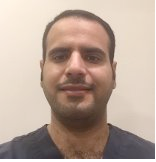
Biography:
Dr. Abdulhameed G.Albeshr earned his BDS degree from King Saud University in Riyadh, SaudirnArabia in 2010. In 2015, he completed his master of public health in community andrnenvironmental health in King Saud bin Abdulaziz University for Health Sciences in Riyadh, SaudirnArabia.
Abstract:
Introduction: Periodontal disease is more prominent in developing countries. It is common in all ages and affect both genders. Unhealthy periodontium cause serious problems for the individual and lead to tooth loss Objective: Determine prevalence of periodontal disease and its risk factors. Method: Electronic search conducted on PubMed using Inclusion criteria; articles in English about prevalence of periodontal disease and its risk factors from 1990 to 2014. 47 articles were identified initially, and after applying exclusion criteria only nine articles were selected for this review. Results: Prevalence of periodontal disease affect 63% - 68%. In the patients above 40 years old, an average of 76% of extracted teeth was due to periodontal disease. The main risk factor for periodontal disease is poor oral hygiene. The role of smoking in developing of periodontal disease was clear. Diabetic patients have the highest prevalence of periodontal disease compared with other diseases, which is 21%. Conclusion: The prevalence of the periodontal disease is high and more studies needed in different cities and in rural areas. Proper sampling and longitudinal studies should be considered to ensure representativeness and confirm the causality. The direct relation between poor oral hygiene and periodontal disease is clear. This review provides convincing evidence about effect of tobacco smoking on oral health. The review shows high prevalence of periodontal disease among diabetic patients. Better understand the full extent and characteristics of periodontal disease in our population beside public health programs will diminish the effects of the disease.
Y.Alhabdan
King Saud bin Abdulaziz University for Health Sciences
Title: Dental Caries Among Children and its Risk Factors; A systematic Review
Time : 15:25-15:45
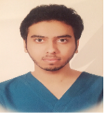
Biography:
Dr. Abdulhameed G.Albeshr earned his BDS degree from King Saud University in Riyadh, SaudirnArabia in 2010. In 2015, he completed his master of public health in community andrnenvironmental health in King Saud bin Abdulaziz University for Health Sciences in Riyadh, SaudirnArabia.
Abstract:
Introduction: Dental caries is one of the most common diseases and most cause of tooth loss in young people. It is painful and can harm nutrition and overall health. However, having baseline information about oral health status is important for planning and evaluation of the improvement in oral health. Objective: Determine prevalence of dental caries among children and identify the associated risk factors. Method: An electronic search conducted to identify articles in PubMed that met our inclusion criteria. A total of 43 articles were identified initially, and after applying exclusion criteria a total of ten articles were selected for this systematic review. Results: Prevalence of caries in primary dentation of children under 6 years ranged from 73-99% and the mean dmft score ranged from 2.92–6.53. In the permanent dentition, the prevalence varied from 87.9-99% and the mean DMFT ranged from 0.78 to 5.94. Prevalence of caries in primary teeth was higher in the urban population. §Poor oral hygiene, dental health knowledge, frequency consumption of cariogenic food, developmental enamel defect and the use of some medication which affect salivary flow rate were found as possible risk factors. A strong possible link was found between poor oral hygiene and prevalence of dental caries with a prevalence of 57.8%. Conclusion: Dental caries is a serious dental public health problem that warrants the immediate attention. Oral health education programs must be deployed and school based approaches should be combined with family and community preventive programs. However, dental caries is both curable and preventable. Therefore, dental caries should be given the top priority and the full resources. Biography Dr. Yazeed A. Alhabdan earned his BDS degree from King Saud University in Riyadh, Saudi Arabia in 2010. In 2015, he completed his master of public health in community and environmental health in King Saud bin Abdulaziz University for Health Sciences in Riyadh, Saudi Arabia.
- Preventive and operative dentistry
Session Introduction
Nilufer Balcioglu
Istanbul University, Istanbul, Turkey
Title: Current status of platelet rich fibrin in oral implantology
Time : 15:45-16:05

Biography:
Dr. Nilufer Balcioglu was graduated from Istanbul University, Faculty of Dentistry in 2001. She obtained the PhD degree from Istanbul University Faculty of Dentistry, Department of Oral Implantology in 2008. She has been working as an Associate Professor at the same department.
Abstract:
First introduced in 2001, Platelet-rich fibrin (PRF) is an autologous fibrin matrix which can be defined as a second-generation platelet concentrate since it does contain leukocytes and does not necessarily require an anticoagulant. Last decades, platelet concentrates have been used in many fields of dentistry. These preparations are aimed to use on operated or wounded sites to achieve a better process of stimulating and accelerate healing mechanism. Although platelet concentrates offer impressive therapeutic perspectives, their clinical relevance implicates a high controversy. Both the translational research and and clinical studies on PRF are a confused and contradictory in a way. The differences mainly depend on preparation techniques and research designs as well. Today several techniques for platelet concentrates are available. The cutting edge technique is Platelet rich fibrin (PRF). PRF is classified as a leukocyte and fibrin concentrate. The main difference between PRF and other platelet concentrates is that the PRF technique does not require any anticoagulant, carrier or activator. PRF can be used successfully solely or in combination with graft materials in the treatment of intrabony periodontal defect, sinüs lifting surgery, socket preservation and peri-implant defects. The goal of this presentation is to describe the current types of state of the art PRF techniques as well as the clinical success rate of these techniques in oral implantology.
Elie E Daou
Lebanese University, Lebanon
Title: Bonding mechanism of porcelain to zirconia frameworks: Problems and solutions?
Time : 16:20 to 16:40
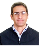
Biography:
Dr. Elie E DAOU was graduated as Dental surgeon in 1994, from Saint Joseph University in Beirut. Then he got a Master in Sciences in 1999, a DES in Prosthodontics in 2002 from the same university, and a UD in “Fundamentals in Medical Research†from F-MRI –LU in 2015. Besides his private practice, he was in Department of Removable Prosthodontics till 2005 at saint Joseph University. He is a Chief Clinical instructor in the Department of Prosthodontics, at the Lebanese University (LU) in Beirut. He is preparing a PhD at the LU. He has published several papers in peer-reviewed journals.
Abstract:
Abstract: Different restorative materials are proposed to the clinician. Their reliability depends on the percentage of restorations still functioning after placement. Different study conditions make the comparison of obtained data challenging. Zirconia frameworks are now widely used by dentists. However, some problems and concerns are reported from the dental market. Zirconia restorations emerged as a substitute to metal ceramic restoration, in response to patients’ esthetics growing demand. The metal-porcelain bonding mechanism is well known; whereas, the zirconia-porcelain interface is still not fully understood. Several factors have been pointed to explain the high porcelain chipping incidence. Latest findings will be detailed in an attempt to explain the zirconia-porcelain bonding. Similarities and differences between metal and zirconia have been raised in the literature.
Narjiss Akerzoul
Consultation Center of Dental Treatments of Rabat,Morroco
Title: Oral Squamous Cell Carcinoma in Pediatric Moroccan Population: A Retrospective Study
Time : 16:40-17:00

Biography:
2005-2011 : Doctorate of Dental Surgery (DDS), Faculty of Dentistry of Rabat, Mohammed V University, Moroccorn2011-2012 : General pratitionner Dentist in Oral Health Center of Guelmim City, Moroccorn2012-Present : 3rd Year Resident Oral Surgeon in Center of Consultation of Dental Treatments of Rabat, Faculty of Dentistry of Rabat, Mohammed V University, Moroccorn2014-2015 : Universitary Diploma of Biostatistics and Research Methodologyrn2014-Present : rn-Author of many International Publications in the field of Oral Surgery and Oral Oncology.rn- International Presenter (Oral & Poster Presenter) in different meetings of Oral Surgery and Head & Neck Oncology in Turkey and the USA.rnMay 2015-Present : Editorial Board Member in Department of Oral and Maxillofacial Surgery of the International Journal of Oral Health and Medical Research ( IJOHMR)rnJuly 2015-Present : Reviewer in OMICS GROUP Biomedical Journals.rnSeptember 2015-Present : Editorial Board Member in Journal of Cosmetology and Orofacial Surgery.
Abstract:
PURPOSE: rnStudy the epidemiological and histopathological profile of the oral cancers in children in care at the Pediatric Hemato-Oncology, Stomatology and Maxillofacial Surgery departments at the 20 August hospital in Casablanca, and at the hospital of children in Rabat. Our aim is to define the importance of the pediatric oncology, especially the one affecting the oral cavity, and also to describe the oral cancers in children, theirs frequences and theirs histopathological characteristics. We did collect 71 patients consultation records and files between 2004- 2012. rnMATERIALS AND METHODS: rnThis is a retrospective study of 126 children hospitalized between 2010 and 2013 of the Pediatric Hemato-Oncology Department, Stomatology and Maxillofacial Surgery department at the 20 August hospital in Casablanca, and also in the Pediatric Hemato-Oncology Department at the hospital of children in Rabat, and in which we did diagnose a confirmed cancer of the oral cavity. rnRESULTS:rn In our sample, all age groups were affected by the disease process, but ages between [0-4] years and between [13-16] were the most affected with an average of age of 8 years, and extremes ranging from 4 months to 16 years. In our population sex sample, we noted a slight female predominance with 50.7% of cases . Non-Hodgkin lymphoma Burkitt was the most common histological type with 35.2% of cases. Cheeks represented the most frequent localization with 37.9% of cases, while the maxillary represented 19.7% of cases. Chemotherapy has been the exclusive therapeutic strategy most used in our sample in 67.6% of cases.rn CONCLUSION: rnEpidemiological, clinical and pathological characteristics of cancers of the oral cavity in our population are not different from the literature data. However, the parents lack of awareness and late diagnosis of these lesions appear to be responsible for the dramatic profile of oral cancers.rn

