Day 2 :
Keynote Forum
Ahmed Halim Ayoub
President of Egyptian society of oral implantology
Keynote: 4D Concept and Immediate Implant Placement
Time : 10:00-10:30

Biography:
Director Dental smile training and educational centrernPresident of Egyptian society of oral implantology Fellow of Seville university,SpainrnPrivate practice limited to dental implants Clinical advisor of Dooox GermanrnDental academy Board member of International group of oral rehabilitation,Francern
Abstract:
4D Concept and Immediate Implant PlacementrnAbstract:rnThe 4th additional axis of time is added to the traditional 3D axes which makes it different to identify the time of extraction and implant placement either immediate implant placement , early implant placement or delayed implant placement.rnThis review focus on each technique and the ideal choice of each technique in every casernIntroduction:rnImmediate implant placement after extraction has become a favored treatment protocol with many clinicians worldwide. There are many advantages to this protocol, amongst them; shortened treatment time, placement of the implant in sound bone that constitutes the socket wall, placement trajectory guidance by the socket and preservation of bone volume. This literature review describes the 4th dimension in implant placement which is the timing of placement after extraction which is a very important factor in immediate placement success rate.rnLearning objectives:rnAfter the presentation the audience shall understand:rn1- The 4th dimension in implant placementrn2- The proper way achieve an perfect immediate implantrn3- Decide whether to place or not to place the implant after extractionrn4- Manage the jumping gaprn5- The proper way to achieve the maximum esthetic outcome in immediate placement.
Keynote Forum
Dr.Omesh Modgill
specialty doctor in the oral surgery department of Kings College Hospital ,London
Keynote: Title: The use of coronectomy in the management of mandibular teeth in paediatric patients

Biography:
Omesh Modgill completed his BDS at the University of Bristol and has since obtained a postgraduate teaching qualification in dental education. Following a year of employment in general dental practice he completed two years of training as a senior house officer in an oral and maxillofacial surgery unit. He currently works as a specialty doctor in the oral surgery department of Kings College Hospital in London. Omesh has previously been published in a leading British dental journal and is actively involved in departmental research and audit projects.
Abstract:
IntroductionrnOccasionally paediatric patients present with symptomatic, unerupted non-third molar mandibular teeth which require surgical intervention but are known to be closely related to adjacent sensory nerves. rnrnCoronectomy is a conservative surgical technique in which the crown of a tooth is removed whilst the roots are deliberately left in situ and may represent the treatment of choice in this situation. Coronectomy is widely and successfully used to reduce the risk of postoperative neuropathy in the surgical management of symptomatic mandibular third molars which are intimately related to the inferior alveolar nerve canal. rnrnMaterials and MethodsrnWe present three paediatric patients who had coronectomy as part of a paediatric dental, orthodontic and oral surgery multidisciplinary treatment plan. They were all assessed clinically, radiographically and with the use of cone beam tomography prior to treatment under day case general anaesthetic. rnrnResultsrnNo patient experienced temporary or permanent postoperative sensory neuropathy or required further surgical intervention for root retrieval following coronectomy.rnrnAn excellent result was achieved for the patient who had orthodontic treatment after coronectomy; the deliberately retained roots had little adverse effect on the alignment of adjacent teeth.rnrnDiscussionrnWe have demonstrated that coronectomy can be used successfully in paediatric patients as an alternative to extraction in the management of mandibular teeth in cases where extraction is considered to present a ‘high risk’ of postsurgical neuropathy. Whilst coronectomy may reduce the risk of neuropathy compared to extraction, it is not a risk or complication- free procedure.rn
Keynote Forum
SAMEH SAMY ABDOU
COURSE DIRECTOR EGYPTIAN SOCIETY OF DENTAL IMPLANT
Keynote: KEYS TO Success for IMPLANTS PLACEMENT WITH IMMEDIATE LOADING
Time : 10:30-11:00

Biography:
Alexandria University Egypt. Diploma in implantology, sevilla university Spain.(2007) consultant in prosthodontics&implantology(2002)private practice limited to prosthodontics & implantology alexandria Egypt . Continuing dental implant course, royal college of surgeon, Edinburg 2005 .dental implants courses Seville university Spain 2006, 2007 .dental education programs ,new York university 2006 . speaker & consultant implantology courses alexandria dental syndicate 2008 ,speaker Egyptian association of dental implant 2009.speaker dgzi congress ,Damascus 2009.speaker Syrian dental association congress 2009,2010speaker Lebanese dental association congress Tripoli 2010 ,2013.speaker sevilla dental implantology congress 2010 .speaker Tanta university dental implant course 2013. Course director Egyptian society of dental implant since 2009.speaker several national & intentional events.
Abstract:
The introduction of osseointegrated implants in dentistry represents a turning point in dental practice.rnThe concept of immediate loading has recently become popular due to less trauma, reduced treatment time, high patient acceptance and better function and esthetics.rnA careful case selection, proper treatment plan, meticulous surgery and proper design of prosthesis are essential for optimal outcomes when this approach is adopted.
Keynote Forum
DR AMOL S. PATIL
Professor /Post-graduate Guide and PhD Guide at Bharati Vidyapeeth University Dental College & Hospital
Keynote: Genetic and biochemical changes in the mandibular condylar cartilage as a function of administration of growth factors and mandibular advancement

Biography:
Currently working as Professor /Post-graduate Guide and PhD Guide at Bharati Vidyapeeth University Dental College & Hospital, Pune. He is one of the few who has completed his PhD in Orthodontics. He was a Gold Medalist in MDS Orthodontics. He has a keen interest in research because of which he pursued his PhD and has total 18 publications out of which 10 are international with high impact factor. He is Reviewer in 17 international journals including AJO, EJO, WJO etc and on board of advisors of 4 international journals. He has been an invited speaker to various international conferences related to basic sciences (1st World molecular and cell biology conference, USA, 2012;Cell Biology Conference, China,2014; Epigenetics and Biotechnology Conference 2014) and recently presented a research paper at 115th AAO conference, San Fransisco. He has presented various papers in national and international conferences for which he has been awarded the best paper awards too. He has keen interest in growth, genetics and basic research.
Abstract:
Growth of the mandibular condylar cartilage (MCC) has always been an area of keen interest for the orthodontists, especially the adaptation of the MCC to forward mandibular positioning. Mandibular forward positioning solicits cellular and molecular responses in the temporo-mandibular joint of growing and non growing animals leading to condylar growth. In contrast, few reports have demonstrated that such adaptive responses to forward mandibular positioning were negligible or non-existent in non-growing animals. It is of importance that our clinical treatment is based on sound scientific understanding of the tissue responses to different treatment modalities. Thus, this presentation will focus on the genetic and biochemical changes in the MCC in young as well as adult rabbits with mandibular advancement. Growth factors are peptides that stimulate cellular growth, differentiation and proliferation. Role of growth factors on the mandibular condylar cartilage has not been investigated thoroughly. Thus, the administration of growth factors with and without mandibular advancement in young and adult rabbits and its influence on condylar growth at the genetic (Vascular endothelial growth factor, SOX-9, Decorin, Matrix gla protein, Matrix mettalo-proteinases), biochemical (Proteoglycans) and histologic level will be discussed.
Keynote Forum
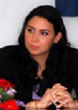
Dr.NARJISS AKERZOUL
Mohammed V University of Rabat, School of Dental Medicine, Rabat-Morocco
Keynote: The Decompression Technique : A Minimally Invasive Oral Surgery Approach
Biography:
Presenter and corresponding Author : Dr NARJISS AKERZOUL rnrn2005-2011 : Doctorate of Dental Surgery (DDS), Faculty of Dentistry of Rabat, Mohammed V University, Moroccorn2011-2012 : General pratitionner Dentist in Oral Health Center of Guelmim City, Moroccorn2012-Present : 3rd Year Resident Oral Surgeon in Center of Consultation of Dental Treatments of Rabat, Faculty of Dentistry of Rabat, Mohammed V University, Moroccorn2014-2015 : Universitary Diploma of Biostatistics and Research Methodologyrn2014-Present : rn-Author of many International Publications in the field of Oral Surgery and Oral Oncology.rn- International Presenter (Oral & Poster Presenter) in different meetings of Oral Surgery and Head & Neck Oncology in Turkey and the USA.rnMay 2015-Present : Editorial Board Member in Department of Oral and Maxillofacial Surgery of the International Journal of Oral Health and Medical Research ( IJOHMR)rnJuly 2015-Present : Reviewer in OMICS GROUP Biomedical Journals.rnSeptember2015-Present : Editorial Board Member and Reviewer in the “Journal of Cosmetology and Orofacial Surgery”. (JCOFS)rn
Abstract:
The purpose of this paper is to present case report of a dentigerous cyst associated to permanent teeth in children treated by conservative techniques.rn Dentigerous cyst is the most common developmental cysts of the jaws. Conservative treatment is very effective to this entity and aims at eliminating the cystic tissue and preserving the permanent tooth involved in the pathology. Two techniques are described as conservative treatment for these cysts, marsupialization and the decompression. rnAn eight years female child was affected by a large lesion at the right side of the mandible associated to tooth 45. The dentigerous cyst promoted severe tooth displacement. rnThe patient was treated with decompression which could manage enough space to do a surgical Orthodontic traction, and therefore place conveniently the permanent tooth.rn
Keynote Forum
Huseyin Avni Balcioglu
Istanbul University Faculty of Dentistry, Istanbul, Turkey
Keynote: Anatomical Sciences and Pediatric Dentistry
Time : 11:00-11:30

Biography:
H. A. Balcioglu, DDS, PhD, holds the position of Associate Professor in the Department of Anatomy at Istanbul University Faculty of Dentistry. After receiving his dental degree from Istanbul University Faculty of Dentistry, in 2001, Dr. Balcioglu worked as a Research Assistant in the department of Anatomy, for five years, and completed the PhD program, while he also worked in private dental practice. He took part in administrative activites associated with his roles as a faculty board member. He is currently a member of the Board of Medical Specialties Reporting System Commission (TUKMOS) of Turkish Ministry of Health. Dr. Balcioglu has lectured as an invited speaker in different symposiums, particularly about TMJ/TMD. His research interests relate primarily to TMJ/TMD, radiologic anatomy and anatomy education.
Abstract:
Intimate knowledge and understanding of the anatomical sciences is paramount in the safe and complete performance of pediatric dentistry. To achieve better results in dental procedures in children and to avoid complications during these procedures, it is essential to establish the location and course of the anatomical structures, particularly the anatomical variations, in the region. There is always a probability of the existence of an anatomical variation that may be developmental, congenital or acquired. Misdiagnosing or overlooking these kind of anatomical variations may lead to neurovascular complications during or after the procedures such as orthodontic corrections, regional anesthesia, implant placement and surgical correction of jaw deformities in children. Besides, knowledge of variational anatomy provides superiority in radiologic interpretation and oral rehabilitation. The training of pediatric dentistry should include a thorough understanding of how the anatomy lectures should be given. This presentation aims to discuss if enough value has been being put on the importance of anatomy in pediatric dentistry training, and if pediatric dentists’ performances rely on a sound anatomy knowledge. Additionally, the presentation will try to refer the audience think on what should be the limits of anatomy course and how should it be taught through the training of pediatric dentistry.
Keynote Forum
Dr.SANTOSH KUMAR
Associate Professor,Department of Orthodontics ,Kothiwal Dental College and Research Centre
Keynote: Effect of cervical pull headgear on head position

Biography:
Santosh Kumar is experienced in treating up to 10-15 patients in a single day. He is involved in the treatment of patients from the age group of 8 to 50 years: Adolescent and adult orthodontics interviewing parents (in case of child patient), patients and identifying their particular problems. He is also experienced in evaluating patient with TMJ complaint, diagnosis and treatment of temporo-mandibular disorders and in the usage of porcelain brackets. He is also involved in undergraduate training from second year BDS onwards, which involves a didactic, practical and clinical component. He is taking lecture classes for undergraduate students and teaching art of basic wire bending
Abstract:
Introduction: Head posture has a significant influence on the carnio-facial morphology. In extended head position, increased facial height, reduced sagittal dimensions and a steeper mandibular plane angle are generally observed, whereas when the head is flexed in relation to the cervical column there is shorter anterior facial height, larger sagittal jaw dimension and a less steep inclination of the mandible. Cervical pull headgear (CHG) is often used to redirect the maxillary growth in Class II malocclusion. Orthopedic force of significant duration is applied on the cervical region during the use of CHG which may have effect on the head posture.rnrnObjective: To evaluate changes in head position following the use of CHG and compare these changes with an untreated control group.rnrnSubjects & Methods: The test group comprised pre-treatment and post-treatment lateral cephalograms of 30 males, aged 11±1.5 years, who were receiving CHG therapy for correction of Class II malocclusion. Pre-observation and post-observation lateral cephalograms of 25 untreated male subjects, aged 11±1.6 years, served as controls. The average treatment time for the treatment group was 12±2.02 months and the average observation period for the control group was 11±1.03 months. Four postural variables (NSL/CVT, NSL/OPT, CVT/HOR, OPT/HOR) were measured to evaluate the head position in all subjects pre- and post-observations.rnrnResults: There was no significant difference in all the measurements concerning the head position within each group (p>0.05). The mean differences of pre- and post-observations of four postural variables in the CHG group were 1.43, 0.9, -1.13, and -1.08, while those of the control group were 1.56, -0.32, -0.24 and 0.04 respectively. There was no significant difference between the headgear and control groups for any of the postural variables measured (p=0.924, 0.338, 0.448 and 0.398 respectively).rnrnConclusions: Although postural variables showed considerable variability in both groups, head position exhibited no significant changes over a period of 11-12 months either in the control or headgear group. This study attempts to clarify to the clinicians that there is no deleterious effect on the head posture with the application of orthopedic force.rn
Keynote Forum
Dr Hafiz Taha
Resident Orthodontics, Section of Dentistry, Department of Surgery, The Aga Khan University Hospital
Keynote: The Association between the Frontal Sinus Morphological Variations and the Cervical Vertebral Maturation for the Assessment of Skeletal Maturity

Biography:
Abstract:
Introduction: The assessment of skeletal maturity is important for planning dentofacial orthopedics or orthognathic surgery for the treatment of different skeletal malocclusions. Cervical vertebral maturation is widely used method to evaluate skeletal maturity of patients undergoing orthodontic treatment. In the past decade, another method is being proposed which is based on frontal sinus morphology. So, the aim of this study is to evaluate the association between frontal sinus morphological variations and cervical vertebral maturation for the assessment of skeletal maturity. rnMethod: Lateral cephalograms of 252 subjects aged 8-21 years were collected from the dental clinics of AKUH. The sample was divided into six groups based on cervical vertebral maturation stages. The frontal sinus index was calculated by dividing frontal sinus height and width and the cervical vertebral maturation stages were evaluated on the same radiograph. Data were analyzed using SPSS (version 19). Kruskal-Wallis test was applied to compare frontal sinus index at different cervical stages and Post hoc Dunnett t3 test was applied to compare frontal sinus index between adjacent cervical stage intervals in males and females. A p-value of ≤ 0.05 was considered as statistically significant.rnResults: The frontal sinus height and width were significantly associated with the individual cervical vertebral maturation stages in males and females. However, frontal sinus index wasn’t significantly associated with the individual cervical vertebral maturation stages in males and females. rnConclusion: Frontal sinus index cannot differentiate between pre-pubertal, pubertal and post-pubertal adolescent growth stages therefore; it cannot be used as a reliable maturity indicator. rn
Keynote Forum
DR. MOHAMMED HUSSEIN AL-BODBAIJ
BDS (KSU), MSC, OMFS (UCL), MFD RCSI
Keynote: Intra-lesional steroid treatment of Central Giant Cell Granuloma of the mandible

Biography:
Abstract:
Central giant cell granuloma (CGCG) is a benign lesion, CGCG occurs mainly in children and young adults with more than 60% of all cases occurring before the age of 30 years and female to male ratio of 2:1. The mandibular / maxillary ratio is from 2:1 to 3:1. rnSurgery is the traditional treatment of CGCG. Calcitonin and intralesional steroid were used with good results. rnIn this case report, a 14 years old Saudi girl presented with a hard swelling of left side of the mandible with few months duration. Investigations including blood tests, radiographs and biopsy were done which confirmed the diagnosed of CGCG. rnLesion has been treated using six weekly intralesional injections of steroid which gave very good result. rnPatient has been followed up for 10 months with radiographic evidence of defect refill with bone and no sign of recurrence.rn
- Advanced research in pediatric dentistry
Session Introduction
Dr.ALESSANDRA MAJORANA
University of Brescia, Italy
Title: Clinical efficacy of a medical device in the treatment of chemotherapy-induced oral mucositis in children
Time : 11:45-12:05

Biography:
Full Professor of Oral Medicine in School of Dentistry of the University of Brescia \r\n1985 Medicine and Surgery Degree (MD)\r\n1990 Odontostomatology Specialization Diploma (DDS)\r\n1991-92 Professor in charge of Oral medicine in School of Dentistry of the University of Brescia\r\n1992 Doctor Researcher in School of Dentistry of the University of Brescia\r\nFrom 1993 Dean of the Department of Oral Medicine and Pediatric Dentistry of Dental Clinic of the Spedali Civili of Brescia \r\n1993-94 Lecturer in Oral Medicine in School of Dentistry of the University of Brescia\r\nfrom 1995 Professor of Pediatric Dentistry in School of dentistry of University of Brescia and Dental Hygiene School\r\nfrom 2001 Full Professor of Oral Medicine and Pediatric Dentistry in School of Dentistry of the University of Brescia \r\nProfessor of dentistry in Medicine Degree Course of University of Brescia\r\n\r\nCounselor of Italian Society of Pediatric Dentistry (SIOI) from 1998 to 2007\r\nPresident of Italian Society of Pediatric Dentistry ( SIOI ) from 2007 to 2010\r\nCounselor of Italian Society of Oral Medicine ( SIPMO) from 2000\r\nMember of’European Academy of Oral Medicine (EAOM)\r\nMember of Mucosite Study Group MASCC\r\n\r\nMember of the National Observatory for the Prevention of Oral Health WHO\r\nCollaborator of the Italian Ministry of Health ( expert in the National Guide Linesfor Oral Health in childhood)\r\n
Abstract:
• Introduction: Oral mucositis (OM) is a severe side effect of anti-cancer therapy, especially in children. It causes a painful inflammatory process, which may have a detrimental effect on quality of life and on therapeutic protocols.\r\nObjectives: The aim was to assess the efficacy of a medical device (Mucosyte ®), respect of placebo, in the treatment of chemotherapy-induced OM in childhood. \r\nMethods: Patients between 5 and 18 years of age undergoing chemotherapy for malignancies diseases with OM grade 1 or 2 were enrolled in this study. They were randomized in group A (treated with Mucosyte ®, 3 mouthwashes/day per 8 days) and group B (treated with placebo, that is an inert water based solution, same dosage). \r\nThe OM scoring was performed at day 1 (diagnosis of OM-T0), after three days of treatment (T1), and at day 8 (T2). Pain was evaluated through the Visual Analogue Scale (VAS) with the same timing of OM measurement. A statistical analysis was performed. \r\nResults: A total of 59 patients were included (28 patients per group). Group A experienced a statistically significative decline of OM just at T2 (p=0.0038) while a statistically significative difference in pain reduction between two groups both at T1 and at T2 (p<0.005) was observed. \r\nConclusions: The present trial demonstrated the efficacy of this medical device (Mucosyte ®) on the treatment of chemotherapy-induced OM in children; in fact, thanks to its barrier effect, it is useful in reducing pain, OM score, burning and erythema. \r\n
Dr.Omesh Modgill
Kings College Hospital ,London
Title: The use of coronectomy in the management of mandibular teeth in paediatric patients
Time : 12:05-12:25

Biography:
Omesh Modgill completed his BDS at the University of Bristol and has since obtained a postgraduate teaching qualification in dental education. Following a year of employment in general dental practice he completed two years of training as a senior house officer in an oral and maxillofacial surgery unit. He currently works as a specialty doctor in the oral surgery department of Kings College Hospital in London. Omesh has previously been published in a leading British dental journal and is actively involved in departmental research and audit projects.
Abstract:
IntroductionrnOccasionally paediatric patients present with symptomatic, unerupted non-third molar mandibular teeth which require surgical intervention but are known to be closely related to adjacent sensory nerves. rnrnCoronectomy is a conservative surgical technique in which the crown of a tooth is removed whilst the roots are deliberately left in situ and may represent the treatment of choice in this situation. Coronectomy is widely and successfully used to reduce the risk of postoperative neuropathy in the surgical management of symptomatic mandibular third molars which are intimately related to the inferior alveolar nerve canal. rnrnMaterials and MethodsrnWe present three paediatric patients who had coronectomy as part of a paediatric dental, orthodontic and oral surgery multidisciplinary treatment plan. They were all assessed clinically, radiographically and with the use of cone beam tomography prior to treatment under day case general anaesthetic. rnrnResultsrnNo patient experienced temporary or permanent postoperative sensory neuropathy or required further surgical intervention for root retrieval following coronectomy.rnrnAn excellent result was achieved for the patient who had orthodontic treatment after coronectomy; the deliberately retained roots had little adverse effect on the alignment of adjacent teeth.rnrnDiscussionrnWe have demonstrated that coronectomy can be used successfully in paediatric patients as an alternative to extraction in the management of mandibular teeth in cases where extraction is considered to present a ‘high risk’ of postsurgical neuropathy. Whilst coronectomy may reduce the risk of neuropathy compared to extraction, it is not a risk or complication- free procedure.rn
Carletto-Körber
Facultad de OdontologÃa, Universidad Nacional de Córdoba
Title: Study of the Population genetic structure and demographic history of Streptococcus mutans

Biography:
Dra. Carletto-Körber Fabiana Marina Pia PhD, Assistant Professor of Child and Adolescent Integral Chair, Faculty of Dentistry, National University of Cordoba. Doctoral and Postdoctoral Fellow in the Department of Science and Technology of the National University of Cordoba. Researcher and Director of research projects subsidized by the Ministry of Science and Technology of the National University of Cordoba. Author of national and international scientific publications. It should also be noted that the research group to whom I have received several national and international awards. President of Dental Circle Punilla. Member of the Board of Dentistry School of Cordoba.
Abstract:
To analyze the genetic structure of Streptococcus mutans, through the use of sequences of strains from Argentina, Japan, Thailand and Finland; and estimate the demographic history of the bacteria through Bayesian analysis. Forty strains of S. mutans were recovered from stimulated saliva of children of Córdoba-Argentina. This work was approved by the Ethics Committee. Of each strain, DNA was extracted DNA and we sequenced the genes aroE, lepC, gyrA and gltA. Sequences from Cordoba-Argentina were aligned with those of strains from Japan (n = 89), Thailand (n = 52) and Finland (n = 12). The total DNA matrix consisted of sequences of 193 strains. Statistical analyzes were performed to determine whether there was evidence of clonality or of recombination in the genes of S. mutans. The pairwise FST between countries was estimated and we performed the Bayesian Skyline Plot and Extended Bayesian Skyline Plot analyses to estimate changes in the effective population size in the last 15,000 years. We detected high allelic diversity (137 alleles) and very low nucleotide diversity, only 12 were shared by two or more countries. The FST values between countries were significant. Of the statistical analyses performed, eight supported the existence of recombination. We detected inter-gene recombination and absence of this mechanism at the intra-gen level. A marked increase in the effective population size was detected approximately 7500 and 5000 years, according to the Bayesian Skyline Plot and Extended Bayesian Skyline Plot analyses, respectively. S. mutans present a recombinant type population genetic structure. The demographic analyses support the hypothesis that the bacteria experimented a population expansion in the last 10000 years.
DR. MOHAMMED HUSSEIN AL-BODBAIJ
BDS (KSU), MSC, OMFS (UCL), MFD RCSI
Title: Intra-lesional steroid treatment of Central Giant Cell Granuloma of the mandible
Biography:
Abstract:
Central giant cell granuloma (CGCG) is a benign lesion, CGCG occurs mainly in children and young adults with more than 60% of all cases occurring before the age of 30 years and female to male ratio of 2:1. The mandibular / maxillary ratio is from 2:1 to 3:1. rnSurgery is the traditional treatment of CGCG. Calcitonin and intralesional steroid were used with good results. rnIn this case report, a 14 years old Saudi girl presented with a hard swelling of left side of the mandible with few months duration. Investigations including blood tests, radiographs and biopsy were done which confirmed the diagnosed of CGCG. rnLesion has been treated using six weekly intralesional injections of steroid which gave very good result. rnPatient has been followed up for 10 months with radiographic evidence of defect refill with bone and no sign of recurrence
- Orthodontics
Session Introduction
Saif alarab Mohmmed
UCL Eastman Dental Institute,U.K
Title: Investigation into the effect of sodium hypochlorite irrigant concentration delivered by a syringe on the rate of bacterial biofilm removal from the wall of a simulated root canal model
Time : 12:25-12:45

Biography:
Abstract:
Aim: Irrigation is a crucial step of successful root canal treatment. It has several important functions, including biofilms disruption. Sodium hypochlorite (NaOCl) is the most popular root canal irriganting solution amongst dentists. This study aimed primarily to develop a transparent root canal model to study in situ Enterococcus faecalis biofilm surface removal rate and remaining attached biofilm when using 5.25%, 2.5% NaOCl and water as irrigating solution for 60s. Methodology: A total of thirty root canal models (n = 10 per group) were manufactured using a 3D printing technique. Each models consisted of two longitudinal halves of an 18 mm length simulated root canal with size #30 and taper 0.06. E. faecalis biofilms were grown on the apical 3 mm for 10 days in Brain Heart Infusion broth. Biofilms were stained using crystal violet. The model halves were reassembled, attached to an apparatus and observed under a fluorescence microscope. Nine mL of NaOCl (5.25%, 2.5%) or water (control) were used for 60 seconds and images were captured every second using a camera. The residual biofilm percentages were measured using image analysis software. Results: The difference in residual biofilm between 5.25% and 2.5% NaOCl groups was 3.3% (±0.3) (p = 0.001). The biofilm removal rate for 5.25% group was 0.6% s-1 (±0.02) (p = 0.001). Whilst, it was 0.4% s-1 (±0.02) (p = 0.001) for 2.5% group. Conclusions: The proposed biofilm model provided a viable mean to investigate biofilm removal under microscopy. The concentration of NaOCl had an influence on the removal rate of biofilm.
Alberto De Stefani
University of Padua, Italy
Title: Patient’s skeletal age evaluation using the middle phalanx maturation of the third finger
Time : 12:45-13:05

Biography:
Abstract:
The orthodontist should evaluate carefully patient’s skeletal age to start an interceptive treatment. Different methods are used: Fishman’s hand wrist radiograph (Fishman 1982), cervical vertebral maturation (CVM method) in a lateral radiograph (Baccetti 2002), middle phalanx maturation of the third finger (MPM method) with an endoral radiograph (Perinetti 2014). Both CVM and MPM methods show six stages (CVS and MPS): two prepubertal stages (CVS 1-2 and MPS 1-2), two pubertal stages (CVS 3-4 and MPS 3-4), and two post-pubertal (CVS 5-6 and MPS 5-6). A diagnostic agreement between the stages of the MPM and the CVM was found so MPM can be used complementary or in alternative to CVM. In literature MPM is evaluated on the right hand as reference. A research made by University of Padua shows a difference of development between the right and the left hand in Caucasian patients. Hand preferred by the patient is also evaluated.
Martina Dandrea
University of Padua, Italy
Title: Nanotechnology for the improvement of tribological properties of orthodontic archwires
Time : 13:50-14:10

Biography:
she's working as a dentist in private practice.
Abstract:
More than 60% of the force used to move teeth through an orthodontic fixed appliance is lost as friction force, with possible loss of anchorage, an enhanced risk of root resorption, an increase in treatment time. The use of molybdenum and tungsten disulfide (MoS2 and WS2) nanoparticles - known for their lubricating properties – can improve from a tribological point of view the substrates to which they are applied, providing a possible solution to minimize friction in an orthodontic system. In this study, nickel (Ni) coatings incorporating MoS2 and WS2 nanoparticles are applied by electrodeposition to 0.019 x 0.025 inch orthodontic stainless steel (SS) wires (Ormco, Glendora, CA, USA). Friction produced by in vitro sliding of coated and un-coated SS wires along self-ligating brackets (Damon Q, Ormco, Glendora, CA, USA; In-Ovation R, GAC International, Islandia, NY, USA) is then evaluated with the use of a universal apparatus for mechanical measurements (Instron 4502). This work shows that good results in terms of friction can be obtained coating SS orthodontic wires with Ni film containing MoS2 and WS2 nanoparticles, providing a possible change in orthodontic materials in the next future.
Martina Mason
University of Padua,Italy
Title: Maxillary transverse discrepancy: posture and gait analysis before and after RPE.
Time : 14:10:14:30

Biography:
Abstract:
Introduction: in the last decades the hypothesis that there is a correlation between dental occlusion and posture has been under investigation by a lot of authors, especially because it has been suggested that disorders of the masticatory system can affect body posture as a whole. the present study aimed to evaluate the effects of rapid palatal expansion (rpe) on gait and posture in children with maxillary transverse discrepancy; differences before and after rpe were assessed. Methods: 50 patients were enrolled, age and bmi were respectively: 9.6±2.1 years, 18.1±1.1 kg/m2, divided into 3 groups: 10 control subjects (cs), 20 patients with unilateral posterior crossbite (cbmono), 20 patients with maxillary transverse discrepancy and no crossbite (nocb). The analyses were carried out in three stages. In the first phase after oral examination, every subject underwent gait analysis, romberg test and surface emg. after 2 moth of the end of rpe’ activation it has been executed surface emg. the last phase was 3 month after the removal of the rpe. The test was executed by means of a 6 cameras sterophotogrammetric system (60-120 hz, bts) synchronized with 2 bertec force plates and a novel pedar plantar pressure system. 3dimensional (3d) joint kinematics, center of pressure displacement (cop), plantar pressure distribution during gait and posturographic parameters were estimated. One way anova or kruskalwallis test was performed among the variables in order to compare the 3 group of subjects, paired t-test was performed instead when comparing a and b conditions within the same group of subjects (p<0.05), tamane t2 or bonferroni correction was used where needed. The presence of asymmetries in each population of subjects was also investigated in term of significant differences between left and right side 3d joints kinematics. Results: the posturographic analysis doesn’t revealed significant differences between cbmono, nocb and control groups. The variables of gait analysis are significant before and after treatment. Surface emg shows an increase of muscle’s force after treatment with a delay of the recruitment of masseter and temporal muscles. Conclusion: it seems to be a relation between occlusion and body posture. it is expressed by dynamic posture in cranio-caudal direction.
Giovanni Bruno
University of Padua,Italy
Title: Safe zones, guidelines for miniplates use in orthodontics
Time : 14:30-14:50

Biography:
Abstract:
Nowadays titanium miniplates are used as a reliable source of absolute anchorage for orthodontic and facial orthopedic movement. Miniplates are generally used apical to dental roots in order to not interfere with dental movements. Cortical bone thickness and density are found to be essential to induce a strong primary stability, the most important factor in anchorage success. Since miniplates induce a stronger primary stability than single miniscrew, they are a valid alternative when a heavy load is necessary or when other absolute anchorage devices fail. A recent research made by University of Padua, Department of Orthodontics, investigated onto cortical bone thickness (mm) and density (HU) in relation to age, gender and skeletal Angle class. No differences were found between groups regarding cortical bone thickness (p-value < 0.05). Differences in cortical bone density were found with adult presenting higher values than adolescents in the three classes (p-value < 0.05). Skeletal pattern, on the other hand, has a slight clinical significance on cortical bone density variations.
Sylvain Haba
Centre Médico-Spiritual Tradi koumi Talitha, Guinea
Title: Oral care and traditional practices harmful to the teeth in Guinea, Sierra Leone and Liberia
Time : 14:50-15:10

Biography:
Sylvain HABA got admission to Bachelor in 1984, orientation to school nurses 1985- 1988. 1989- 1997 Internship in a medical post in Sèbètèrè Gaoual Prefecture. 1992-1993 Remote Training on medical semiology. In 1998 admis to test the public service.2004-2005 Training in traditional medicine in DR Congo. Back Guinea in 2006 establishment of the Centre Médico-Spiritual Tradi koumi Talitha (Marc5: 41-42) to (Labe) Guinea.2008 Transfer of the center in Conakry
Abstract:
In Guinea the oral diseases have long been given the importance for health instead tooth brushes plants were used because of their effects, analgesic, anti-inflammatory, antiseptic, antibiotic. 1) Phyllanthus floribunda -family euphorbiacéa 2) Alchornea cordifolia -Family: euphorbiacea 3) the Gongrenema latifolia -family asclépiadacéa 4) Hyppocratéa indica Family: hyppocratacé. Oral infections were treated with honey, citrus juice and medical leaves Euphorbia hirta children, were added sheets phyllanthus floribunda adults. Today modern toothbrushes and toothpaste are used to reduce the risks and complications dough. These are the traditional practices affecting the health dangerously. Weakening the role of the teeth (mechanical digestion), the risk of tetanus and HIV by cutting the teeth. At 20 years my incisors were cut. To conclude my article, I ask the international committee to intervene so that this traditional practice is prohibited. The epidemic of Ebola fever is endemic by harmful traditional practices that do not improve prevention. I ntil the day hui continue the same practices here are the results of my queries in the last two months between Guinea Conakry and Sierra Leone these young people and older people live within 200 km of my capital Conakry. Hardly, they accepted photos for fear of being prosecuted because the area was hard hit by the Ebola fever.
Sameh Samy Abdou
Alexandria University, Egypt
Title: Constraints and obstacles in dental implant treatment
Time : 15:10-15:30

Biography:
Sameh Samy Abdou obtained Diploma in Implantology from Sevilla University, Spain in 2007. He is a Consultant in Prosthodontics & Implantology and his private practice is limited to Prosthodontics & Implantology at Alexandria, Egypt. He continued dental implant course, at Royal College of Surgeon, Edinburg in 2005 and in Seville University, Spain in 2006-2007. He participated in Dental Education Programs in New York University in 2006. He served as a speaker &consultant of implantology courses at Alexandria Dental Syndicate in 2008 and speaker of Egyptian Association of Dental Implant 2009, DGZI congress at Damascus in 2009, Syrian Dental Association Congress in 2009-2010, Lebanese Dental Association Congress at Tripoli in 2010-2013, Sevilla Dental Implantology Congress in 2010 and in Tanta University Dental Implant Course, 2013. He is a Course Director of Egyptian society of dental implant since 2009 and speaker of several national & international events.
Abstract:
The appropriated placed dental implants are the most reliable method of tooth replacement. Keys to dental implant success should include proper case selection, excellent surgical technique, placing adequate restoration on the implant, educating the patient to maintain meticulous oral hygiene. Although the overall success rate of dental implant is very high, there are few absolute Constraints and Obstacles. The experienced trained clinician should have little difficulty in assessing patients as suitable.
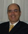
Fabio Savastano
International College of Neuromuscular Orthodontics and Gnathology, Italy
Title: What to do for TMD in children: a neuromuscular perspective."
Time : 15:45-17:15
Biography:
Graduated in Medicine and Surgery in 1987 cum laude at the University of Naples, Italy. Master in Orthodontics at the University of Padua in 1990. Adjunct professor at the University des Les Valls, Andorra. Practice limited to orthodontics and gnathology since 1991 in Albenga. President of ICNOG, International College of Neuromuscular Orthodontics and Gnathology, and International member of the AAO, American Association of Orthodontics, is member of numerous associations and has lectured in Brazil, Canada,U.A.E., Spain,Bahrain,India and Italy on "Neuromuscular Orthodontics".
Abstract:
Neuromuscular dentistry is the understanding of the relationship between the Temperomandibular joints(TMJ), teeth, muscles and nerves. It enables the optimum physiologic position of the jaw to be established to assist in the correction of the underlying causes of craniofacial – Temperomandibular joint, head and neck pain. Neuromuscular dentistry is also used to determine the optimum physiologic jaw position prior to complex dental restorative procedures, cosmetic dentistry, dental sleep medicine procedures, dentofacialorthopaedics and orthodontics. It is a treatment modality of dentistry that focuses on correcting the physiologic “misalignment†of the jaw at the Temperomandibular joint (TMJ). TMJ disorders are not a problem limited to adults. This lecture focuses on how to diagnose and treat TMD in children and young adults. Neuromuscular-functional orthodontics is an easy error free procedure that will take the practitioner to treat TMJ disfunction in children during interceptive therapy.


