Day 1 :
Keynote Forum
T P Chiang
Canada China Child Health Foundation, Canada
Keynote: Importance of Dentistry in the management of cleft lip/palate
Time : 09:25-10:00
Biography:
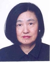
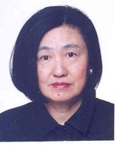
T P Chiang is the President of Canada China Child Health Foundation, Canada. She has done her BSc, DDS and Doctorate in Dental Medicine from Dalhousie University, Canada. She has completed her Master of Science in Epidemiology from Harvard University. She did her Post-doctoral studies at Massachusetts Institute of Technology, Boston Children's Hospital in Pediatric Dentistry. She is a Professor at University of British Columbia, Beijing Children's Hospital, Beijing Institute of Pediatrics and Children's Hospital of Harbin. She is the Honorary President of Nanjing Medical University Dental Hospital; Consultant at child health hospitals of Guangzhou, Suzhou Health College, and Chongqing Medical University.
Abstract:
Dentistry plays an important role in the management of cleft lip and palate. Pediatric dentist is generally the first person to have contact with the cleft child patient. It is a common congenital maxillofacial deformity involving serious tissue defects and associated problems, early treatment would allow better prognosis in the end result of cleft lip/palate treatment. There is profound tissue defect involvement with maxillary bone segment loss and tissue displacement, affecting both physical appearance and function. This deformity causes major challenges because of associated problems, i.e. feeding, conduct disorder, high treatment cost, ear infection, hearing loss, language difficulty. Prevalence of cleft lip and palate ranks extremely high among congenital anomalies in the whole world. It is the second most common birth defect in the United States. Worldwide, oral clefts in any form occur in about one in every 700 live births. In fact prevalence rate vary among different countries and even ethnic groups within the same country. Overall, higher rates have been reported in Asians and American Indians (1/500 births), and lower rates have been reported in African-derived populations (1/2,500 births). Current surgical techniques and managements greatly improve the treatment effectiveness of cleft lip/palate. Cleft lip and palate management extend beyond simple surgical repair and encompass restoration of appearance and function, psychological problem, and changes in growth and development. An integrated and multidisciplinary approach is particularly important in achieving optimal result and is almost standard in US and Canada. Collaborative team would involve: pediatric dentist, plastic surgeon, anesthesiologist, orthodontist, maxillofacial surgeon, and speech pathologist, audiologist, feeding nurse, pediatrician and otolaryngologist. Cleft sequential treatment approaches growth stages with different therapeutic targets. Neonatal period pursue physical appearance/ functionality; prepubertal period guide arch form development and completion of alveolar bone graft; puberty aims at improve function; orthognathic surgery follows growth and development completion. There are four stages of treatment approaches: neonatal phase-obturation, tissue molding and orthopedic appliance; primary and mixed dentition phase-orthodontics and orthopedics; permanent dentition-comprehensive orthodontics and orthopedics; and final phase-+/- orthognathic surgery including distraction osteogenesis. The goals of the treatment protocol are to perform pre-surgical orthopedics during infancy including selection/insertion of pre-surgical device, followed by surgical repair of cleft lip/palate. This will be followed by expansion/alignment, alveolar bone graft closure of oronasal fistula during the mixed dentition phase, fixed orthodontics during the permanent dentition phase synchronized with orthognathic surgery. This will be finalized with prosthetic and esthetic reconstruction. These dental treatments would complement the rest of the interdisciplinary team. This would enable the team to achieve maximum result in the comprehensive patient management allowing the building of proper dental arch form, facial and dental alignment, speech, esthetics, function and psychological aspects of the patient.
Keynote Forum
Reem Hanna
King’s College Hospital NHS Foundation Trust, UK
Keynote: The advantages of carbon dioxide laser applications in paediatric oral surgery: A prospective cohort study
Time : 10:00-10:35
Biography:
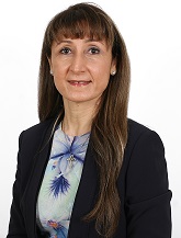
Hanna R is an Associate Specialist in Oral Surgery at King’s College Hospital. She is a Registered Specialist in Oral Surgery in UK. She is honorary Senior Lecture at UCL Eastman Dental Institute where she leads the fellowship in laser dentistry and advanced oral surgery courses. She was appointed as Visiting Professor in Surgical Sciences and Integrated Diagnostics Department, University of Genoa (UNIGE) in 2015. She is a faculty member who teaches Master of Science students in laser dentistry program at UNIGE. She lectures nationally and internationally on the applications of oral laser therapy. Her great interest is utilizing photobiomodulation in tissue regeneration and neuropathic pain.
Abstract:
The aim of this study is to evaluate and demonstrate the advantages of the carbon dioxide laser in paediatric oral surgery patients in terms of less post-operative complications, healing without scaring, functional benefits, positive patient perception and acceptance of the treatment. 100 fit and healthy paediatric patients (aged 4–15 years) were recruited to undergo laser surgery for different soft tissue conditions. The outcome of these laser treatments was examined. The Wong-Baker Faces Pain Rating Scale (Fig. 1) was employed to evaluate the pain before, immediately after laser treatment in the clinic and one day after post-operatively at home. Post-operative complications and patients’ perception and satisfaction were self-reported during a review telephone call the day after treatment. The patients were reviewed two weeks after surgery. Laser parameter was 1.62 W, measured by power meter, continuous wave mode with 50% emission cycle. The beam spot size at the target tissue was 0.8 mm. The pain score pre-operative during and immediately after laser treatment was rated 0. While the pain scores one day after surgery were rated between 0 and 2, the healing time was measured over two weeks. None of the patients reported post-operative complications after surgery. Patients’ perception and acceptance were rated very well. Laser dentistry is a promising field in modern minimally invasive dentistry, which enables provision of better care for children and adolescents. In this cohort study, the use of the carbon dioxide laser therapy offers a desirable, acceptable and minimally invasive technique in the surgical management of soft tissues in paediatric oral surgery with minimal post-operative complications.
Recent Publications
- Suter V G, Altermatt H J, Sendi P and Mettraux G (2010) CO2 and diode laser for excisional biopsies of oral mucosal lesions. A pilot study evaluating clinical and histopathological parameters. Schweiz Monatsschr Zahnmed 120(8):664–7.
- Puthussery T, Shekar K and Gulati K (2011) Use of carbon dioxide 
laser in lingual frenectomy. J Oral Maxillofac Surg 49:580–581.
- Pié-Sánchez J, España-Tost A-J, Arnabat-Domínguez J and Gay-Escoda C (2012) Comparative study of upper lip frenectomy with the CO2 laser versus the Er, Cr: YSGG laser. Med Oral Patol Oral Cir Bucal 17(2):228–23.
- Vescovi P, Corcione L, Meleti M, Merigo E, Fornaini C, Manfredi M, Bonanini M, Govoni P, Rocca J R and Nammour S (2010) Nd:YAG laser versus traditional scalpel. A preliminary histological analysis of specimens from the human oral mucosa. Lasers Med Sci 25(5):685–91.
- Puthussery T, Shekar K and Gulati K (2011) Use of carbon dioxide laser in lingual frenectomy. J Oral Maxillofac Surg 49:580–581.
Keynote Forum
Abdullah Mohammed Alzahem
King Saud bin Abdulaziz University for Health Sciences, Saudi Arabia
Keynote: Cheek-bite keratosis among temporomandibular disorders patients: An important diagnostic sign
Time : 11:00-11:35
Biography:
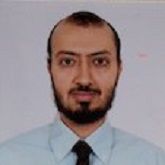
Abdullah Mohammed Alzahem is an Assistant Professor in King Saud bin Abdulaziz University of Medical Education. He completed his BDS in 1995; postgraduate studies in Temporomandibular Joint Disorders and Advanced General Dentistry in USA. He was awarded the prestigious Fellowship of General Dentistry Academy (FAGD) in 2004, and in the same year appointed as Dental Consultant. He completed successfully two-year Master program in Medical Education (MME) in 2009. He completed his PhD degree in Medical Education at Erasmus University Rotterdam, and was appointed as Director of Quality Assurance in King Saud bin Abdulaziz University for Health Sciences.
Abstract:
Introduction: Cheek-biting commonly reported by patients with Temporomandibular Disorders (TMDs). This check biting may cause cheek-bite keratosis. This research aim is to study the prevalence of cheek-bite keratosis among TMDs patients.
Materials & Methods: Cross-sectional survey conducted on 373 TMDs patients seen in the TMJ clinic by one TMJ specialist since 2013. Convenient sampling technique was followed where all screened patients having TMDs included in the study.
Results: TMDs patients who have check-bite keratosis are 226 patients (60.6%). Female TMDs patients are the majority (75.60%) and 78.8% of TMDs patients with cheek bit keratosis were female. The highest number of TMDs patients (4.6%) was at age of 20 years old.
Conclusion: Cheek-bite keratosis is an important sign for TMDs screening for the general dentist in the first dental visit. Dentist who find cheek-bite keratosis during intra-oral examination, advised to ask more screening questions and do more clinical examination for TMDs.
- Special Session
Location: Johnson
Session Introduction
Ziad E F Noujeim,
Lebanese University, Lebanon
Title: Management of dental pathologies and odontogenic cysts and tumors of the jaws in pediatric patients: Our experience
Time : 11:35-12:20
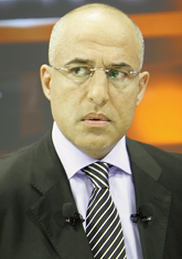
Biography:
Ziad Noujeim is Senior Lecturer and Clinical Professor of Oral Surgery at Lebanese University, Beirut. He is attending Oral Surgeon at Lebanese Army Hospital and Baabda University Hospital. He is Diplomate of European Board of Oral Surgery, Fellow of International College of Dentists and American College of Oral and Maxillofacial Surgeons, and former Clinical Fellow at Massachusetts General Hospital, Harvard School of Dental Medicine in Boston, USA. He has published 24 articles in reputed journals. He is presently Editor-in-Chief of the Journal of the Lebanese Dental Association and Section Editor of the Annals of Maxillofacial Surgery.
Abstract:
Few oral surgeons have extensive experience in the management of dental and jaw pathologies in children and adolescents. Treatment of such rare pathologies is usually multidisciplinary, but the main core is often surgical, yet not always followed by reconstructive surgery. In addition to establishing a sound histopathologic diagnosis, it is important to understand and evaluate the biological behavior of such pathologies; some of these, despite being labeled as benign, are quite aggressive clinically and can cause significant bone loss and impairment if not diagnosed early and adequately treated. Pediatric odontogenic jaw cysts and tumors are unusual, and most of them are asymptomatic, the most common lesions being the dentigerous cysts and the keratocystic odontogenic tumor. It is of utmost importance that pediatric dentists, orthodontists, and general practitioners should be familiar with these three groups of lesions which can manifest as jaw swelling, delays in dental eruptions, facial asymmetry, displacement of teeth, alteration in occlusion and loosening of associated or adjacent teeth. In our presentation, we will address a case series of pediatric dental irregularities (impacted teeth and teeth-like structures) and odontogenic cysts and tumors. All these pathologies required surgical intervention, including surgical extraction, biopsy, curettage, ostectomy, enucleation and chemoablation.
- Oral Implantology| Public Health Dentistry |Pediatric Dentistry | Endodontics | Orthodontics & Dental Implants | Periodontics
Location: Johnson
Chair
T P Chiang
Canada China Child Health Foundation, Canada
Co-Chair
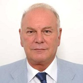
Francesco Inchingolo
University of Bari, Italy
Session Introduction
Gustavo Vicentis de Oliveira Fernandes
Salgado de Oliveira University, Brazil
Title: The use of Biomaterials in oral rehabilitation: New trends
Time : 13:20-13:45

Biography:
Gustavo Vicentis de Oliveira Fernandes is a Dentist. He has completed his Master’s degree in Medical Science, and PhD in Dentistry. He has his expertise in Periodontics, Implantology and Oral rehabilitation. He is a Researcher in biomaterials, oral implants and tissue reconstruction. He is a Full Professor in Periodontics (Salgado de Oliveira University, Brazil). He has a passion in rehabilitate patients, improving the aesthetic and wellbeing. His work is based in scientific evaluation with clinical application.
Abstract:
Biomaterials revolutionized the dynamics of treatments in the area of medicine and dentistry, enabling critical tissue corrections and achieving mimicry. In this large group named biomaterials, the gold standard appears, known as autogenous, which has several growth factors involved and many favorable properties, which helps a lot in the regenerative process. Therefore, rehabilitating patients today has become more accessible and sometimes more challenging, since the professional must know which technique to use, which material to choose, and have satisfactory manual detraining to achieve the desired success. Thus, the objective of this lecture will be to present tissue reconstructions with basic and advanced techniques and techniques focused on periodontics and implantology. Finally, we can observe that the new techniques and materials, such as the collected blood, favored the treatments significantly in dentistry, making possible extreme cases.
Recent Publications
-
Costa NMF, Yassuda DH, Sader MS, Fernandes GVO, Soares GA and Granjeiro JM (2015). Osteogenic Effect of Tricalcium Phosphate Substituted by Magnesium with Genderm® Membrane in Rat Calvarial Defect Model. Materials Science and Engineering C 61.
-
Sena LA, Almeida MSM, Fernandes GVO, Guerra R, Castro-Silva II, Granjeiro JM and Achete CA (2014). Biocompatibility of wollastonite-poly (N -butyl-2-cyanoacrylate) composites. Journal of Biomedical Materials Research Part B Applied Biomaterials 102(6).
-
Fernandes GVO, Cavagis ADM, Ferreira CV, Olej B, Leão MS, Yano CL, Peppelenbosch M, Granjeiro JM and Zambuzzi WF(2013). Osteoblast Adhesion Dynamics: A Possible Role for ROS and LMWâ€PTP. Journal of Cellular Biochemistry 115(6):1063-1069.
-
Aline Muniz de Oliveira AM, Castroâ€Silva II, Fernandes GVO, Melo BR, Alves ATNN, Júnior AS, Lima ICB and Granjeiro JM (2013). Effectiveness and acceleration of bone repair in criticalâ€sized rat calvarial defects using lowâ€level laser therapy. Laser in Surgery and Medicine. 46(1):61-67.
Cherif Massoud
Crystalign, Lebanon
Title: The roles of artificial intelligence and technology in orthodontic treatments today
Time : 13:45-14:10

Biography:
Cherif Massoud is a key opinion leader in orthodontic aligners because of his combined expertise in orthodontics on one hand and 3d printing modeling and design on the other. He is the founder and CEO of Crystalign.
Abstract:
Braces are from the past. The science of orthodontics is going through a rebirth with the advancement of technology, artificial intelligence and machine learning. 3D printed invisible aligners are being used to treat severe orthodontic cases. We will present the latest techniques that are pushing the traditional boundaries.
Key words: Aligners, 3d printing, digital workflow, Crystalign
Mohammad Reza Salahi
Shiraz University of Medical Science, Iran
Title: Comparison of dental and periodontal problems in liver and renal failure patients with normal population
Time : 14:10-14:35

Biography:
Mohammad Reza Salahi (DDS) was born on December 26th 1984 in Shiraz (IRAN). He completed his graduation from Shiraz medical university in Shiraz (IRAN) IN 2012. He got fellowship in implant surgery in 2016. He is working on some articles about laser in Endodontics.
Abstract:
Transplantation is the best treatment for organ failure. However, the patients who have to undergo transplantation should wait for a long time, and this might contribute to developing some complications. Moreover, after excessive search in medical journals, we found that previous studies mostly have focused on the oral cavity in the transplant patients, in chronic renal failure and in liver diseases. However, few studies evaluate dental health status and radiographic evaluation in liver failure or renal failure patients. This motivated us to conduct the present study to compare the renal failure and liver failure patients with normal people with regard to the findings of oral cavity and oral plain radiography. This is a descriptive study conducted on the patients with chronic renal or liver failure were registered for the transplantation on the waiting list, in Nemazee hospital transplant center. Oral examinations and oral plain graphy was requested. Having consulted the statistic professor, we assigned the participants to three groups: choronic renal failure CRF (n: 50), liver disease (n: 50), and normal group (n: 50). The software SPSS was used for data analysis. Findings: The three groups participated in the present study were the normal group (n: 50), CRF group (n: 50), and liver failure (n: 50) group. Gingival recession was observed in 16, 23, and 33 patients in Normal, CRF group, and liver failure group respectively (Pvalue<0.05). We also noticed bone loss in 13, 23, and 29 patients in normal group, CRF, liver failure group (Pvalue: 0.002). The mean number for missing teeth needed extraction and root canal therapy, the teeth for which root canal therapy was performed the teeth were filled before the study, and the teeth in need of filling for the liver failure group was more than that in the normal group. (The Pvalue was <0.05), however, the mean for CRF group was significantly lower than that for the normal group (Pvalue<0.05). Compared with the normal group the mean number for missing teeth in CRD group was significantly higher. Gingival recession and bone loss in liver failure and chronic kidney disease group were significantly more than that in the normal group. The prevalence of caries was lower in CRF patients compared with the normal group; in contrast, missing teeth, filled teeth. Dental filling and root canal therapy were lower in the liver failure patients than that in the normal group. Missing teeth was more prevalent in chronic kidney disease than normal group.
Recent Publications
1. Sagheb MM, Sharifian M, Ahmadi S, Moini M, Rais-Jalali GA, Behzadi S, Roozbeh J, Jalaian H, Nikeghbalian S, Bahador A, Salahi H, Salehipoor M, Kazemi K and Malek-Hosseini SA (2011). Comparison of immediate renal dysfunction in split and partial liver transplantation versus full size liver transplantation in Shiraz transplant centre. Ann Transplant; 16(2):36-42.
- Jamali R, Khonsari M, Merat S, Khoshnia M, Jafari E, Bahram Kalhori A, Abolghasemi H, Amini S, Maghsoudlu M, Deyhim MR, Rezvan H and Pourshams A(2008). Persistent alanine aminotransferase elevation among the general Iranian population: prevalence and causes World J Gastroenterol 14(18):2867-71.
- Sheehy EC, Roberts GJ, Beighton D and O'Brien G (2000).Oral health in children undergoing liver transplantation. Int J Paediatr Dent 10(2):109-19.
- de la Rosa-Garcia E, Mondragon-Padilla A, Irigoyen-Camacho ME and Bustamante-Ramirez MA (2005). Oral lesions in a group of kidney transplant patients. Med Oral Patol Oral Cir Bucal 10(3):196-204.
- Grubbs V, Gregorich SE, Perez-Stable EJ and Hsu CY (2009) Health literacy and access to kidney transplantation Clin J Am Soc Nephrol 4(1):195-200.
Yasmeen A AlHaizan
King Saud University, Saudi Arabia
Title: Psychological stress and burnout among healthcare workers in Department of Dental Services, MNGHA (Ministry of National Guard - Health Affairs): A cross sectional survey
Time : 14:35-15:00

Biography:
Yasmeen A AlHaizan is pursuing her Bachelor of Dental Surgery (BDS) at College of Dentistry, King Saud University, Saudi Arabia. In 2017, she participated in 9th Research Summer School held at King Abdullah International Medical Research Center (KAIMRC), which grew her interest and developed her skills in research.
Abstract:
Introduction: Professional burnout, a prolonged response to stress, would possibly affect the standards of patient care. Burnout is defined as emotional exhaustion, depersonalization, and diminished personal accomplishment.
Aim: To identify and compare psychological stress and burnout levels among different job titles and specialties in dental department, MNGHA. Also, to determine the effect of marital status, age, and gender on stress and burnout levels.
Methods: Convenient sampling approach was used to distribute the questionnaire in the dental department, MNGHA (n=177, response rate=88.5%). Two validated questionnaires, psychological stress measure-9 (PSM-9) and Maslach Burnout Inventory–Human Services Survey (MBI-HSS), were used.
Results: Mean level (and standard deviation) of stress was 32.60 (11.43), with the highest stress levels seen in consultants and residents (39.17% and 38.33%). Hygienists and technicians scored the highest lack of personal accomplishment (24.53%), consultants scored the highest emotional exhaustion (24.64%), while residents scored the highest impersonal response toward patients (26.67%).
Conclusion: Participants with the job title consultant and resident are shown to be the most stressed and burnt-out category among the dental department. Specialty, gender, age and marital status are not shown to be risk factors in our study. Stress and burnout should be reduced to maintain the standards of patient care.
Huda Salem Alrakaf
Prince Sultan Military Medical City, KSA
Title: Oral histopathological changes in premature infants
Time : 15:00-15:25

Biography:
Huda Salem Alrakaf completed her Graduation from King Saud University (KSU), Riyadh, Saudi Arabia. She then obtained her Master of Science in Dentistry at KSU. In Madrid, Spain 2003, she earned her specialized certificate on high risk and special need children. Later in the year 2007, she took a course in psychopathology of children and intervention at Howard University, Washington DC, USA. Her achievements in 2001 were remarkable she did the first publication of both intra-nasal midazolan in conscious sedation of young pediatric dental patients and dental management for coach syndrome.
Abstract:
Preterm and low birth weight children comprise approximately 10% of all live births. It is an enormous global problem that is exacting a huge loss emotionally, physically and financially on families along with medical systems premature children experience many oral complications associated with their preterm birth. Prematurely born infants have short prenatal developmental period and they are prone to many serious medical problems during the neonatal period which may affect the development of oral tissue. It was reported that premature born whom were intubated had developed erosions of the maxillary anterior alveolar ridge. The dragging motion of the orotracheal tube traumatized the mucosa with consequent ulceration and pressure necrosis of the alveolar ridge and underlying tooth buds. Hence, premature neonates require assisted ventilation using nasotracheal or orotracheal tubes. However, orotracheal intubation is not free of complication. Histopathological changes to the airway, mucosa, damage to the larynx, subglottic and bronchial dentofacial deformities (primary tooth dilaceration cross bites), poor speech intelligibility. Enamel alteration (enamel hypoplasia, enamel opacities), dental size (small primary tooth crown size associated with BW should be considered studies of tooth size in all population) and changes to the dental mineral content. At the same time oral lesions represent a wide range of diseases often creating apprehension and anxiety among parents. Early examination and prompt diagnosis can aid in prudent management and serves as baseline in the future course of the disease. This present review is on the deleterious effect of preterm birth and oral tube on oral structures and their development. Implications for long term care and follow up of dental concerns are also discussed.
- Pediatric Dentistry| Operative Dentistry | Restorative Dentistry | Dental Case Reports | Endodontics
Location: Johnson
Chair
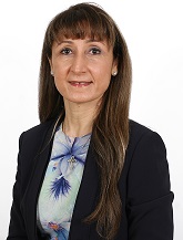
Reem Hanna
King’s College Hospital NHS Foundation Trust, UK
Co-Chair
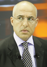
Ziad E F Noujeim
Lebanese University, Lebanon
Session Introduction
Umber Zahra
Lahore Medical and Dental College, Pakistan
Title: Esthetic rehabilitation of an 8-year-old patient with non-syndromic oligodontia and the role of multidisciplinary team: A case report and a literature review
Time : 15:40-16:05

Biography:
Umber Zahra has completed her Bachelor of Dental Surgery (BDS) from Institute of Dentistry, CMH Lahore Medical and Dental College, Pakistan. Her work experience includes practicing part time in a General Practice. She chose to specialize in Operative Dentistry to gain the knowledge and skills to manage complex esthetic, restorative and endodontics cases of both children and adults. She is enrolled in the Residency Program (Operative Dentistry) at Lahore Medical and Dental College to become a Fellow of College of Physicians and Surgeons, Pakistan. She is currently working as a Post-graduate Resident and Clinical Demonstrator in the Deparmtent of Operative Dentistry, Lahore Medical and Dental College. Pakistan.
Abstract:
Oligodontia is the congenital absence of six or more than six teeth in either permanent or primary dentition. Because of the missing teeth in these patients esthetic, functional and psychological problems may arise. This article reports a rare case of non-syndromic oligodontia in an eight year old female patient of Asian origin. A total number of seven permanent and six primary anterior teeth were congentially absent. There are very few cases reported in literature in which primary teeth are congenitally missing. Psychological stress due to missing teeth was evident in the patient’s behavior. A ‘fixed anterior esthetic space maintainer’ was fabricated and fixed into the patients mouth. Conditions like oligodontia have a substantial impact on the functional and psychological maturation of the child. Management of conditions could be challenging, the key for a successful outcome is early diagnosis and proper treatment planning involving all other concerned specialties i.e. through multidisciplinary approach. The rehabilitation with an interim prosthesis, like an esthetic space maintainer or removable partial dentures, at an early age and later with osseointegrated implants has shown promising results from the functional and psychological point of view for such cases.
Edmond Koyess
Lebanese University, Lebanon
Title: Postoperative pain after the use of waveone gold files or protaper next files system in shaping the root canal: A randomized controlled clinical trial
Time : 16:05-16:30
Biography:
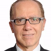
Edmond Koyess completed his graduation from Saint-Joseph University in Beirut Lebanon. He received Post-graduate certificate in Oral Biology and Endodontics from Rene Descartes, Paris. He completed his Doctorate degree in Odontologic Sciences from Lebanese University. Presently, he is Director of Master in Endodontics at Lebanese University and Fellow of International College of Dentists. He is an International Lecturer and Opinion Leader in Endodontics.
Abstract:
Postoperative pain after shaping and cleaning the canal system can vary in degree of occurrence. The most commonly described cause of postoperative pain is the presence in the root canal system and the eventual extrusion of microorganisms inevitable in addition to pushing debris of contaminated dentin, necrotic pulp tissue and. An inflammation can follow this phenomenon accompanied by acute pain due to local pressure and oedema that triggers a postoperative pain of different levels. Modern NiTi files are widely used in dental offices as well as limited practices to Endodontics. The kinematics of NiTi files are of two types: continuous rotation and reciprocating motions. Debris forcing outside the canal can be variable according to the dynamics of the shaping files whether rotating or reciprocating. Protocol of pre-enlargement as recommended by the manufacturer and the shaping procedure being comparable, it can be accepted that pain following the shaping procedures can be assessed in a randomized clinical study.
In this presentation the author will expose the study conducted in a private clinic with limited practice of endodontics by the same operator having at least two years experience using experience in both systems thus, comparing the incidence of post-instrumentation pain associated with both NiTi files system following canal shaping and cleaning. To assess the effect of shaping the canal solely without the effect of obturation, the procedures were conducted in two visits. At the second appointment patients were asked to rate the intensity of pre-instrumentation and post-instrumentation pain (at 2, 4, 6, 8, 24, 48 h) using the VAS score.
Maya Awada
Barts and The London School of Medicine and Dentistry, London
Title: Dentists in the forensic field
Time : 16:30-16:55

Biography:
Maya Awada is a dentist who graduated from the Lebanese University-School of Dental Medicine. She has an interest in forensic science and decided to advance her knowledge in this field. She is a master student in Forensic Medical Science at Barts and The London School of Medicine and Dentistry- Queen Mary University of London.
Abstract:
Forensic odontology or forensic dentistry is the implementation of dental evidence in the legal and criminal field. It involves the application of dental knowledge mainly in the scope of human identification both in ordinary cases of individual identification and in disaster victim identification (DVI), examination of patterned injuries caused by teeth that are frequent in sexual crimes and child abuse cases and age assessment all in the interest of justice. The uniqueness of an individual dentition its resistance to temperature and time are the major reasons behind odontology being a key factor in solving forensic cases. The presentation will offer an overview of forensic odontology, the process of identification using dental records and dental imaging, the importance of this method in mass disasters such as Thailand tsunami, and the concept of bite mark analysis.
Key words: forensic dentistry, identification, victim, bite mark analysis, disaster victim identification.
Mohamad Saad El Masri
University College London, London
Title: Process of establishing the academic and clinical aspect of SCD in the Arab World, particularly Lebanon: A promising challenge
Time : 16:55-17:20

Biography:
Mohamad Saad El Masri is specialised in Special Care Dentistry (Dentistry for people with disability). His aim is to trigger the establishment of the Clinical and Academic aspect of SCD across the MENA countries-particularly Lebanon, to enable the delivery of oral healthcare for people with an impairment or disability.
Abstract:
The Arab region of the world is rapidly changing and advancing. There are striking differences from the developed world in terms of prevalence and type of diseases leading to various forms of disability. Furthermore, political challenges, financial constraints, limited healthcare systems, and negative attitudes and beliefs towards individuals with disability are all factors that can influence the provision of healthcare services for these individuals. These factors can additionally impact on access to oral healthcare, which is often not considered a priority.
Although approximately 15% of the population in the Arab states are estimated to be living with disability. Attitudes toward disability in this region have been found to be negative. There are few studies in the region with regards to the dental needs of individuals with disabilities. Dental education and training to provide oral health care for patients with disability in the Arab world are remarkably limited. Countries in the region should take advantage from the available evidence provided by the International Association for Disability and Oral Health (iADH) and the British Society for Disability and Oral Health (BSDH) and consider establishing SCD within the undergraduate curriculum.
The aims of the presentation are to review the impact of disability across the region, identify factors that may influence access to healthcare, review barriers in order to enable the imminent approach to address the provision of SCD, and outline the pathway that will enable the establishment of the Academic and Clinical Aspect of SCD in the Arab world, and particularly in Lebanon.
- Workshop
Location: Johnson
Session Introduction
Ziad E F Noujeim
Lebanese University, Lebanon
Title: Problem-based learning-PBL: A Dentistry trend for today’s lifestyle
Time : 11:30-13:15
Biography:
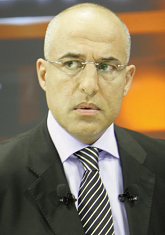
Ziad E F Noujeim is a Senior Lecturer, Postgraduate Tutor, and Clinical Professor of Oral Surgery and Oral Pathology at Lebanese University, Beirut. He is an attending oral surgeon at Lebanese Army Hospital and Baabda University Hospital. He is a Diplomate of the European Board of Oral Surgery, Fellow of the International College of Dentists and the American College of Oral and Maxillofacial Surgeon, and former Clinical Fellow at Massachusetts General Hospital/Harvard School of Dental Medicine, in Boston, USA. He has published 22 scientific articles in reputed journals, among them, 11 were listed on PubMed. He is presently an Editor-in-Chief of the Journal of the Lebanese Dental Association-JLDA and Section Editor of the Annals of Maxillofacial Surgery-AMS.
Abstract:
Over the past three decades, many dental schools, worldwide, have introduced Problem-Based Learning-PBL-in their programs. One of the main reasons for change has included the obvious dissatisfaction of dental students with the conventional model of dental education. PBL was introduced in its modern form at McMaster University Medical School in Canada, in the 1960s, and as applied now, the problem comes first, before any formal study or relevant literature review: this learning method is student-centered, i.e., students are directly involved in deciding what and how they will learn. In this tutorial-like learning, students are guided by a facilitator, and the learning takes place in relatively small groups. PBL contrasts with conventional educational approaches to dental education that are usually teacher-centered rather than student-centered. This method fulfills three important principles related to the development of new knowledge, namely, activation of prior knowledge, elaboration of knowledge, and encoding specificity. It also improves clinical and diagnostic reasoning ability, and fosters development of skills and attributes that oral health professionals will need in the future. PBL tutorial identifies cues in the dental problem presentation, formulates a coherent statement about it, generates hypotheses that arise out of the cues, decides on an inquiry plan, and discusses the final treatment planning and its implementation. Our specialized PBL workshop will focus on dental analgesia difficulties, dental and oral diseases, and jaw disorders. During 120 minutes, this method will help participants integrating knowledge with daily dental practice, nurtures their ability to analyze dental problems, and develops teamwork and communication skills in their minds. Ultimately, PBL meets dentistry trends for today’s lifestyle by cultivating mind independence, scientific curiosity, and necessary skills for self-directed, life-long learning. With PBL applied in dentistry, oral health providers are able to provide the highest standards of dental care in a proficient, professional, caring, and ethical manner.
Recent Publications
- Abdelkarim A, Schween D and Ford T (2018). Attitudes towards Problem-Based Learning of Faculty Members at 12 U.S. Medical and Dental Schools: A Comparative Study J Dent Educ. 82(2):144-151.
- Chuenjitwongsa S, Oliver RG and Bullock AD (2017). Developing educators of European undergraduate dental students: Towards an agreed curriculum. Eur J Dent Educ.
- Von Bergmann H, Walker J, Dalrymple KR and Shuler CF(2017). Dental Faculty Members' Pedagogic Beliefs and Curriculum Aims in Problem-Based Learning: An Exploratory Study. J Dent Educ. 81(8):937-947.
- Bai X, Zhang X, Wang X, Lu L, Liu Q and Zhou Q (2017). Follow-up assessment of problem-based learning in dental alveolar surgery education: a pilot trial. Int Dent J. 67(3):180-185.
- Hoffman GR and Sasidharan P (2016). A Medical School Elective Can Promote an Interest in and an Exposure to the Scope of Oral and Maxillofacial Surgery: Education, Exemplars, and Electives. J Oral Maxillofac Surg. 74(4):665-7.
- Orthodontics | Pediatric Dentistry| Orofacial Myology |Oral and Maxillofacial Surgery | Public Health Dentistry
Location: Johnson
Chair
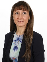
Reem Hanna
King’s College Hospital NHS Foundation Trust, UK
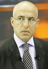
Co-Chair
Ziad E F Noujeim
Lebanese University, Lebanon
Session Introduction
Sanjana Sudarshan
King’s College London, United Kingdom
Title: Audit on recall intervals of high risk children at Wedgwood house dental practice
Time : 14:45-15:10

Biography:
Sanjana Sudarshan is a General Dental Practitioner and is due to commence a Senior House Officer position in Oral and Maxillofacial Surgery at The Royal London Hospital. She is focused on furthering her post graduate education and is on course to gaining The Diploma of Membership of the Joint Dental Faculties at The Royal College of Surgeons of England. Sanjana has a keen interest in Paediatric and Orthodontic Dentistry and aspires to specialise in the field of Orthodontics.
Abstract:
Child caries is a significant issue in our population, in both permanent and deciduous dentition, with at least a third of children across all age groups having caries into dentine. The benefits of frequent oral health reviews are numerous including early detection and enhanced prevention. This is of increased importance for children possessing high caries risk factors such poor plaque control, highly cariogenic diets or children with previous or existing carious lesions. This audit uses retrospective data collection to assess whether appropriate recall intervals are being assigned to high risk paediatric patients at Wedgwood House Dental Practice in accordance to the guidance provided by National Institute of Clinical Excellence (NICE). The first 50 high risk patients under the age of 18 attending for an Oral Health Assessment or Oral Health Review were included in the study sample. The results of the first cycle of the audit showed that only 32% of high risk children were given the appropriate recall interval of 3 months. Furthermore, it was shown that only 56% of high risk children were assessed and assigned the correct risk status. Therefore, interventions to increase the adherence to guidelines were put in place: teaching and training for staff; posters displayed in key locations throughout the practice; handouts of guidance literature; and encouraging dentists to add a reminder of the correct recalls to their proformas. Time was allowed for the interventions to take effect and the audit was repeated using same methodology as previously. The second cycle of audit results collected showed that now 96% of children were being correctly assessed and 60% of these high risk children were now correctly being recalled at 3 months. This shows an 87% increase in correct recall intervals being assigned to high risk children, thus highlighting the positive impact of the interventions.
Key words: Paediatric, caries, risk assessment, oral health review, recall intervals, prevention.
Marwa Sabry
Kafr Elsheikh University, Egypt
Title: Role of dentist in detection and reporting child abuse and neglect
Time : 15:10-15:35

Biography:
Marwa Sabry is an Assistant Lecturer at Paediatric Dentistry and Dental Public Health Department, Kafr El-Sheikh University, Egypt. She has completed a Bachelor of Dentistry degree and MS in Dental Public Health from Alexandria University, MS in Hospital Administration from High Institution of Public Health, and Diploma in Total Quality Management from American University in Cairo. She is working in the field of Health Administration, Quality management and Public Health since 10 year. She plays an active role in community, she cooperates number of national and international associations to provide health educational programs and epidemiological surveys to raise people’s awareness.
Abstract:
Background & Aim: Child abuse and neglect is a very serious problem that has long consequences for those involved and for society in general. Dental professionals are in an exceptional position to identify and report these cases. Statistics of previous studies revealed that only one percent of dentists reported suspected cases. Aims of the study are to help dentists to learn about their ethical responsibilities and legal obligation toward child abuse cases and how to detect and report them.
Methods: This study will demonstrate types and consequences of child abuse and neglect and the prevalence worldwide. In addition, ethical and legal concerns of different health care organizations related to dentists’ are reporting suspected cases. Oral symptoms of child abuse and neglect will be discussed and classified according to the type: physical abuse, sexual abuse and neglect. Physical abuse may results in lacerations of tongue, oral mucosa, palate, gingiva alveolar mucosa or frenum; fractured, displaced, or avulsed teeth; facial bone and jaw fractures; burns; or other injuries. Sexual abuse may represents significant oral manifestations as oral and perioral gonorrhoea, unexplained erythema or petechiae of the palate, particularly at the junction of the hard and soft palate, pseudomembranous and condylomatous lesions of lips, tongue, palate and nose-pharynx. Dental neglect is detected by untreated early Childhood Caries, odontogenous infection or pain, periodontal diseases.
Conclusion: Child abuse and neglect can be prevented by dentist’s awareness about their roles of reporting these cases and strengthen their abilities to detect them in early stage.
Juliana Gabrielle Martins
Federal University of Minas Gerais, Brazil
Title: Hidden sugar in food and the prevalence of dental caries in pre-school children
Time : 15:35-16:00

Biography:
Juliana Gabrielle Martins is a Pediatric Dentist and has completed her MSc and PhD in Pediatric Dentistry from UFMG, Belo Horizonte, Brazil. She was a Visiting Researcher at Harvard University (2015/2016). She has published papers in reputed journals and has been serving as an Editorial Board Member of the Journal of Alcohol and Drug Abuse. Furthermore, she is currently a Reviewer of the Journal of Addiction & Neuropharmacology and for the Journal Ciência & Saúde Coletiva, and she is the Collaborator of the Master in Pediatric Dentistry at Barcelona University and Researcher at Johns Hopkins University - Public Policy Center (UPF).
Abstract:
Increased consumption of processed foods, both by children and adults, is one of the factors linked to the onset of caries since most of them have sugar in their composition. The objective was to analyze the dietary pattern in children attending the "Baby Clinic" of the Universidade Federal de Minas Gerais (UFMG), Brazil and, consequently, to analyze the dental caries experience in early childhood. This is a cross-sectional observational study with a sample of 134 children, aged 0-36 months, attending until the first semester of 2017. The habits of food, hygiene, sociodemographic and economic factors were evaluated by means of a questionnaire addressed to parents / guardians. To evaluate the prevalence of dental caries, oral clinical examination was performed. Descriptive and bivariate analyzes were performed (p<0.05). The prevalence of caries was 17.9% (n = 24). Of the 134 children treated 71 (53.0%) were males, with a mean age of 14 months. It was observed that 70.9% of the mothers who reported not adding sugar to the bottle did so without knowing. The most cited bottle-fed foods were 'Mucilon', 'Toddy' and 'Farinha Lactea Cereal Flour'. In the bivariate analysis, maternal schooling (p <0.01), adding sugar in the bottle (p = 0.04) and consuming sugary foods more than 3 times a day (p <0.01) were associated with presence of dental caries. The high prevalence of dental caries observed in young children is a reality. Therefore, the need for family counseling for food and dental care is observed earlier.
Recent Publications
- Martins J G, Paiva H N, Paiva P P, Ferreira R C, Pordeus I, Zarzar P M and Kawachi I (2017). New Evidence about the "Dark Side" of Social Cohesion in Promoting Binge Drinking among Adolescents. PLOS ONE. https://doi.org/10.1371/journal.pone.0178652.
- Martins-Oliveira J G, Jorge K O, Ferreira R C, Ferreira E F, Vale M P and Zarzar P M (2016).Risk of alcohol dependence: prevalence, related problems and socioeconomic factors. Ciência & Saúde Coletiva, (21) 17-26.
- Video Presentations
Location: Johnson
Session Introduction
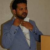
Sudhir Dole
Maharashtra University of Health Sciences, India
Title: Ozonotherapy in Dentistry: A Revolution in Dentistry
Time : 16:15-16:45
Biography:
Sudhir Dole is the First Certified Ozone Therapy Dentist Specialist in India practicing Ozone Therapy in Dentistry at Mumbai, India. He is the Speaker and Mentor for certification courses in ozone dentistry and key note speaker at various conferences in India.
Abstract:
Biological dentistry using synergistic ozone technologies and minimal invasive/non-invasive dentistry with 100% safe protocols and skills make us learn how and when to use ozone, learn which protocol to use and how to adapt it in clinical practice, learn how to motivate both team members and patients, learn what limitations ozone has in clinical care and to know what other areas of healthcare ozone can be used in. Expanding services and investing in ozone technologies get returns of the fees paid in one month, and to become a biological dentist and have a synergistic approach in your practice using ozone. Bioenergetics and bio-oxidative ozone has its effects and treatment functions in oral medicine replacing as a safest adjunct and fastest healer than all other therapies as well can be effective in conjunction with other treatment modalities. The topical application ozone and aqueous ozone which is completely safe is highly recommended as supplementary adjunct and supportive adjunct to other supportive treatments like irrigation system, disinfectants, fumigation, purified water and bottled water, natural antibiotic, immunomodulatory stimulant, anti-inflammatory and analgesic in all specialities of dentistry. Properties of ozone: 3000 times more effective disinfectant because of high concentration of oxygen content and minimal or zero side effects; high antimicrobial activity; anti-inflammatory and analgesic; faster healing capacity and hemostat; induces increased blood circulation; gaseous ozone is highly effective for cure of dental problems as there is easy accessibility to any areas and difficult anatomies; its supplementary benefits for irrigation agents are very useful in endodontics for minimizing the cytotoxicity and it's irritating effects on surrounding structures and to heal its effects on vital structures; disinfection and increasing potency of liquids, increasing efficiency of equipments and instruments by using ozonated water in clinic/dental set up; good carrier for tissues, grafts, membranes, hair strands for maintaining their vitality; increases the natural immunity and improved natural immunity by inducing the immunoglobulins and its activity and; it's benefits to activate RBC, WBCS and blood derivatives which cause increase in blood circulation to high level thus by increasing the energy by promoting carbohydrate and protein metabolisation.
Jyoti Oberoi
Dr D.Y.Patil Dental College and Research Institute, India
Title: Reintroducing hypnosis in Paediatric Dentistry
Time : 16:45-17:15

Biography:
Jyoti Oberoi is pursuing PHD in clinical hypnosis from Dr. D.Y.Patil School of Dentistry, Deemed University, Nerul Navi Mumbai, Maharashtra from where she did her graduation as bachelor of dental surgery (B.D.S.) and Post Graduated in Pedodontics. She is director of Gurukrupa Medical Trust and practicing there as dentist, Pedodontist and Implantologist since 2010. As well she is running ADC Inc. as training centre and academy for dental education. She has presented more than 10 posters/papers in national/international conferences and won prizes for the same. She has 8 publications on her name.
Abstract:
Majority of pediatric dental patients reveal a great anxiety and fear during routine oral procedures. Such attitude of children to dental procedures is a cause of irregular visits to dental clinics, which, in consequence, may lead to greater damage to teeth, which otherwise could have been saved by simple procedures. Clinical hypnosis could be a non−invasive therapeutic option to increase treatment comfort both for the patients and dentists. This article gives an overview about some basic facts and the main indications for hypnosis in dentistry. The indications for using hypnosis in dentistry are: the management of fear and anxiety, hypnosis for dental analgesia, control of bleeding, control of salivation, control of bruxism, control of gag reflex, pediatric dental hypnosis. Commonly used techniques are also listed. This kind of psychotherapy may be used in everyday dental practice, however some profound knowledge in this field is needed from a clinician.
Key words: dental hypnosis, dental fear and anxiety, dental phobia, dental analgesia.


